 Movie
Movie Controller

[English] 日本語
 Yorodumi
- PDB-8rag: Crystal structure of class Ie ribonucleotide reductase R2 subunit... -
+ Open data

- Basic information
| Entry | Database: PDB / ID: 8rag | |||||||||
|---|---|---|---|---|---|---|---|---|---|---|
| Title | Crystal structure of class Ie ribonucleotide reductase R2 subunit without Y150 modification from Gardnerella vaginalis | |||||||||
 Components Components | ribonucleoside-diphosphate reductase | |||||||||
 Keywords Keywords | OXIDOREDUCTASE / Ribonucleotide reductase R2e / Class Ie RNR / ferritin-like superfamily / NrdF / RNR beta-subunit | |||||||||
| Function / homology |  Function and homology information Function and homology informationribonucleoside-diphosphate reductase complex / ribonucleoside-diphosphate reductase / ribonucleoside-diphosphate reductase activity, thioredoxin disulfide as acceptor / deoxyribonucleotide biosynthetic process / metal ion binding Similarity search - Function | |||||||||
| Biological species |  Gardnerella vaginalis ATCC 14019 (bacteria) Gardnerella vaginalis ATCC 14019 (bacteria) | |||||||||
| Method |  X-RAY DIFFRACTION / SYNCHROTRON / MOLECULAR REPLACEMENT / Resolution: 1.9 Å X-RAY DIFFRACTION / SYNCHROTRON / MOLECULAR REPLACEMENT / Resolution: 1.9 Å | |||||||||
 Authors Authors | John, J. / Hogbom, M. | |||||||||
| Funding support |  Sweden, 2items Sweden, 2items
| |||||||||
 Citation Citation | Journal: Commun Biol / Year: 2025 Title: Characterization of a second class Ie ribonucleotide reductase. Authors: John, J. / Lundin, D. / Branca, R.M. / Kumar, R. / Srinivas, V. / Lebrette, H. / Hogbom, M. | |||||||||
| History |
|
- Structure visualization
| Structure viewer | Molecule: MolmilJmol/JSmol |
|---|
- Downloads & links
-Download
| PDBx/mmCIF format | 8rag.cif.gz | 628.6 KB | Display | PDBx/mmCIF format |
|---|---|---|---|---|
| PDB format | pdb8rag.ent.gz | 463.4 KB | Display | PDB format |
| PDBx/mmJSON format | 8rag.json.gz | Tree view | PDBx/mmJSON format | |
| Others |  Other downloads Other downloads |
-Validation report
| Arichive directory | https://data.pdbj.org/pub/pdb/validation_reports/ra/8ragftp://data.pdbj.org/pub/pdb/validation_reports/ra/8rag | HTTPS FTP |
|---|
-Related structure data

-Links
 PDBj
PDBj
- Assembly
| Deposited unit | 
| ||||||||||||
|---|---|---|---|---|---|---|---|---|---|---|---|---|---|
| 1 | 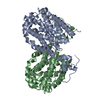
| ||||||||||||
| 2 | 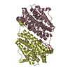
| ||||||||||||
| Unit cell |
|
-Components
| #1: Protein | Mass: 41757.574 Da / Num. of mol.: 4 Source method: isolated from a genetically manipulated source Source: (gene. exp.) Gardnerella vaginalis ATCC 14019 (bacteria)Gene: HMPREF0421_21333 / Production host: References: UniProt: E3D8A3, ribonucleoside-diphosphate reductase #2: Water | ChemComp-HOH / |  Mass: 18.015 Da / Num. of mol.: 1196 / Source method: isolated from a natural source / Formula: H2O Mass: 18.015 Da / Num. of mol.: 1196 / Source method: isolated from a natural source / Formula: H2OHas protein modification | N | |
|---|
-Experimental details
-Experiment
| Experiment | Method: X-RAY DIFFRACTION / Number of used crystals: 1 |
|---|
- Sample preparation
| Crystal | Density Matthews: 4.07 Å3/Da / Density % sol: 69.8 % / Description: transparent rhombohedron |
|---|---|
| Crystal grow | Temperature: 293 K / Method: vapor diffusion, sitting drop / pH: 6.5 Details: Morpheus G4 condition (0.1 M Carboxylic acids, 0.1 M Buffer System 1 pH 6.5, 37.5% v/v Precipitant Mix 4) + 4% tert-butanol |
-Data collection
| Diffraction | Mean temperature: 100 K / Serial crystal experiment: N |
|---|---|
| Diffraction source | Source: SYNCHROTRON / Site: Diamond  / Beamline: I04 / Wavelength: 0.95374 Å / Beamline: I04 / Wavelength: 0.95374 Å |
| Detector | Type: DECTRIS EIGER2 XE 16M / Detector: PIXEL / Date: Jul 8, 2023 |
| Radiation | Protocol: SINGLE WAVELENGTH / Monochromatic (M) / Laue (L): M / Scattering type: x-ray |
| Radiation wavelength | Wavelength: 0.95374 Å / Relative weight: 1 |
| Reflection | Resolution: 1.9→47.74 Å / Num. obs: 214258 / % possible obs: 99.95 % / Redundancy: 21 % / Biso Wilson estimate: 36.95 Å2 / CC1/2: 0.999 / CC star: 1 / Rmerge(I) obs: 0.1704 / Rpim(I) all: 0.03803 / Rrim(I) all: 0.1746 / Net I/σ(I): 12.94 |
| Reflection shell | Resolution: 1.9→1.968 Å / Rmerge(I) obs: 2.239 / Mean I/σ(I) obs: 0.68 / Num. unique obs: 21312 / CC1/2: 0.596 / CC star: 0.864 / Rpim(I) all: 0.4927 / Rrim(I) all: 2.293 |
- Processing
| Software |
| |||||||||||||||||||||||||||||||||||||||||||||||||||||||||||||||||||||||||||||
|---|---|---|---|---|---|---|---|---|---|---|---|---|---|---|---|---|---|---|---|---|---|---|---|---|---|---|---|---|---|---|---|---|---|---|---|---|---|---|---|---|---|---|---|---|---|---|---|---|---|---|---|---|---|---|---|---|---|---|---|---|---|---|---|---|---|---|---|---|---|---|---|---|---|---|---|---|---|---|
| Refinement | Method to determine structure: MOLECULAR REPLACEMENT / Resolution: 1.9→47.74 Å / SU ML: 0.1779 / Cross valid method: FREE R-VALUE / σ(F): 1.34 / Phase error: 21.3556 Stereochemistry target values: GeoStd + Monomer Library + CDL v1.2
| |||||||||||||||||||||||||||||||||||||||||||||||||||||||||||||||||||||||||||||
| Solvent computation | Shrinkage radii: 0.9 Å / VDW probe radii: 1.1 Å / Solvent model: FLAT BULK SOLVENT MODEL | |||||||||||||||||||||||||||||||||||||||||||||||||||||||||||||||||||||||||||||
| Displacement parameters | Biso mean: 42.47 Å2 | |||||||||||||||||||||||||||||||||||||||||||||||||||||||||||||||||||||||||||||
| Refinement step | Cycle: LAST / Resolution: 1.9→47.74 Å
| |||||||||||||||||||||||||||||||||||||||||||||||||||||||||||||||||||||||||||||
| Refine LS restraints |
| |||||||||||||||||||||||||||||||||||||||||||||||||||||||||||||||||||||||||||||
| LS refinement shell |
|
