+ Open data
- Basic information
| Entry | Database: PDB / ID: 8r9v | ||||||||||||||||||
|---|---|---|---|---|---|---|---|---|---|---|---|---|---|---|---|---|---|---|---|
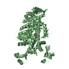
| Title | CryoEM structure of the primed actomyosin-5a complex | ||||||||||||||||||
 Components Components |
| ||||||||||||||||||
 Keywords Keywords | MOTOR PROTEIN / Myosin / Actin / Actomyosin / Primed actomyosin | ||||||||||||||||||
| Function / homology |  Function and homology information Function and homology informationestablishment of endoplasmic reticulum localization to postsynapse / regulation of postsynaptic cytosolic calcium ion concentration / melanosome localization / endoplasmic reticulum localization / locomotion involved in locomotory behavior / melanin metabolic process / vesicle transport along actin filament / unconventional myosin complex / insulin-responsive compartment / developmental pigmentation ...establishment of endoplasmic reticulum localization to postsynapse / regulation of postsynaptic cytosolic calcium ion concentration / melanosome localization / endoplasmic reticulum localization / locomotion involved in locomotory behavior / melanin metabolic process / vesicle transport along actin filament / unconventional myosin complex / insulin-responsive compartment / developmental pigmentation / secretory granule localization / Regulation of MITF-M-dependent genes involved in pigmentation / Regulation of actin dynamics for phagocytic cup formation / hair follicle maturation / actin filament-based movement / melanin biosynthetic process / melanocyte differentiation / actomyosin / melanosome transport / myosin complex / intermediate filament / odontogenesis / long-chain fatty acid biosynthetic process / insulin secretion / cytoskeletal motor activator activity / myosin heavy chain binding / microfilament motor activity / tropomyosin binding / actin filament bundle / troponin I binding / filamentous actin / pigmentation / mesenchyme migration / exocytosis / smooth endoplasmic reticulum / cytoskeletal motor activity / skeletal muscle myofibril / actin filament bundle assembly / striated muscle thin filament / skeletal muscle thin filament assembly / actin monomer binding / photoreceptor outer segment / skeletal muscle fiber development / stress fiber / vesicle-mediated transport / titin binding / actin filament polymerization / myelination / visual perception / secretory granule / protein localization to plasma membrane / actin filament / filopodium / synapse organization / small GTPase binding / Hydrolases; Acting on acid anhydrides; Acting on acid anhydrides to facilitate cellular and subcellular movement / cellular response to insulin stimulus / disordered domain specific binding / calcium-dependent protein binding / melanosome / lamellipodium / actin binding / cell body / chemical synaptic transmission / calmodulin binding / postsynapse / hydrolase activity / protein domain specific binding / neuronal cell body / calcium ion binding / positive regulation of gene expression / glutamatergic synapse / magnesium ion binding / Golgi apparatus / ATP hydrolysis activity / ATP binding / identical protein binding / cytosol / cytoplasm Similarity search - Function | ||||||||||||||||||
| Biological species |   | ||||||||||||||||||
| Method | ELECTRON MICROSCOPY / single particle reconstruction / cryo EM / Resolution: 4.4 Å | ||||||||||||||||||
 Authors Authors | Klebl, D.P. / McMillan, S.N. / Risi, C. / Forgacs, E. / Virok, B. / Atherton, J.L. / Stofella, M. / Winkelmann, D.A. / Sobott, F. / Galkin, V.E. ...Klebl, D.P. / McMillan, S.N. / Risi, C. / Forgacs, E. / Virok, B. / Atherton, J.L. / Stofella, M. / Winkelmann, D.A. / Sobott, F. / Galkin, V.E. / Knight, P.J. / Muench, S.P. / Scarff, C.A. / White, H.D. | ||||||||||||||||||
| Funding support |  United Kingdom, United Kingdom,  United States, 5items United States, 5items
| ||||||||||||||||||
 Citation Citation |  Journal: Nature / Year: 2025 Journal: Nature / Year: 2025Title: Swinging lever mechanism of myosin directly shown by time-resolved cryo-EM. Authors: David P Klebl / Sean N McMillan / Cristina Risi / Eva Forgacs / Betty Virok / Jennifer L Atherton / Sarah A Harris / Michele Stofella / Donald A Winkelmann / Frank Sobott / Vitold E Galkin / ...Authors: David P Klebl / Sean N McMillan / Cristina Risi / Eva Forgacs / Betty Virok / Jennifer L Atherton / Sarah A Harris / Michele Stofella / Donald A Winkelmann / Frank Sobott / Vitold E Galkin / Peter J Knight / Stephen P Muench / Charlotte A Scarff / Howard D White /    Abstract: Myosins produce force and movement in cells through interactions with F-actin. Generation of movement is thought to arise through actin-catalysed conversion of myosin from an ATP-generated primed ...Myosins produce force and movement in cells through interactions with F-actin. Generation of movement is thought to arise through actin-catalysed conversion of myosin from an ATP-generated primed (pre-powerstroke) state to a post-powerstroke state, accompanied by myosin lever swing. However, the initial, primed actomyosin state has never been observed, and the mechanism by which actin catalyses myosin ATPase activity is unclear. Here, to address these issues, we performed time-resolved cryogenic electron microscopy (cryo-EM) of a myosin-5 mutant having slow hydrolysis product release. Primed actomyosin was predominantly captured 10 ms after mixing primed myosin with F-actin, whereas post-powerstroke actomyosin predominated at 120 ms, with no abundant intermediate states detected. For detailed interpretation, cryo-EM maps were fitted with pseudo-atomic models. Small but critical changes accompany the primed motor binding to actin through its lower 50-kDa subdomain, with the actin-binding cleft open and phosphate release prohibited. Amino-terminal actin interactions with myosin promote rotation of the upper 50-kDa subdomain, closing the actin-binding cleft, and enabling phosphate release. The formation of interactions between the upper 50-kDa subdomain and actin creates the strong-binding interface needed for effective force production. The myosin-5 lever swings through 93°, predominantly along the actin axis, with little twisting. The magnitude of lever swing matches the typical step length of myosin-5 along actin. These time-resolved structures demonstrate the swinging lever mechanism, elucidate structural transitions of the power stroke, and resolve decades of conjecture on how myosins generate movement. | ||||||||||||||||||
| History |
|
- Structure visualization
| Structure viewer | Molecule:  Molmil Molmil Jmol/JSmol Jmol/JSmol |
|---|
- Downloads & links
- Download
| PDBx/mmCIF format |  8r9v.cif.gz 8r9v.cif.gz | 335.4 KB | Display |  PDBx/mmCIF format PDBx/mmCIF format |
|---|---|---|---|---|
| PDB format |  pdb8r9v.ent.gz pdb8r9v.ent.gz | 270.7 KB | Display |  PDB format PDB format |
| PDBx/mmJSON format |  8r9v.json.gz 8r9v.json.gz | Tree view |  PDBx/mmJSON format PDBx/mmJSON format | |
| Others |  Other downloads Other downloads |
-Validation report
| Arichive directory |  https://data.pdbj.org/pub/pdb/validation_reports/r9/8r9v https://data.pdbj.org/pub/pdb/validation_reports/r9/8r9v ftp://data.pdbj.org/pub/pdb/validation_reports/r9/8r9v ftp://data.pdbj.org/pub/pdb/validation_reports/r9/8r9v | HTTPS FTP |
|---|
-Related structure data
| Related structure data |  19013MC  8rbfC  8rbgC M: map data used to model this data C: citing same article ( |
|---|---|
| Similar structure data | Similarity search - Function & homology  F&H Search F&H Search |
- Links
Links
- Assembly
| Deposited unit | 
|
|---|---|
| 1 |
|
- Components
| #1: Protein | Mass: 92247.883 Da / Num. of mol.: 1 / Mutation: S217A, Deletion DDEK 594-597 Source method: isolated from a genetically manipulated source Source: (gene. exp.)   Santiria sp. sf9 (plant) / References: UniProt: Q99104 Santiria sp. sf9 (plant) / References: UniProt: Q99104 | ||||||||||
|---|---|---|---|---|---|---|---|---|---|---|---|
| #2: Protein | Mass: 41888.652 Da / Num. of mol.: 3 / Source method: isolated from a natural source / Source: (natural)  References: UniProt: P68135, Hydrolases; Acting on acid anhydrides; Acting on acid anhydrides to facilitate cellular and subcellular movement #3: Chemical | ChemComp-PO4 / | #4: Chemical | ChemComp-MG / #5: Chemical | ChemComp-ADP / Has ligand of interest | Y | Has protein modification | Y | |
-Experimental details
-Experiment
| Experiment | Method: ELECTRON MICROSCOPY |
|---|---|
| EM experiment | Aggregation state: FILAMENT / 3D reconstruction method: single particle reconstruction |
- Sample preparation
| Component |
| |||||||||||||||||||||||||||||||||||
|---|---|---|---|---|---|---|---|---|---|---|---|---|---|---|---|---|---|---|---|---|---|---|---|---|---|---|---|---|---|---|---|---|---|---|---|---|
| Source (natural) |
| |||||||||||||||||||||||||||||||||||
| Source (recombinant) |
| |||||||||||||||||||||||||||||||||||
| Buffer solution | pH: 7 | |||||||||||||||||||||||||||||||||||
| Buffer component |
| |||||||||||||||||||||||||||||||||||
| Specimen | Embedding applied: NO / Shadowing applied: NO / Staining applied: NO / Vitrification applied: YES Details: Actin-myosin mixture comprising 13 uM myosin (pre-mixed with ATP) and 13 uM actin | |||||||||||||||||||||||||||||||||||
| Specimen support | Details: 300 mesh copper grids with copper nano-wires (SPT Labtech) Grid material: COPPER / Grid mesh size: 300 divisions/in. / Grid type: Quantifoil | |||||||||||||||||||||||||||||||||||
| Vitrification | Instrument: HOMEMADE PLUNGER / Cryogen name: ETHANE / Humidity: 60 % / Chamber temperature: 293 K Details: F-actin was mixed with myosin-5a (S1 1 IQ motif, residues 1-797, S217A, DDEK 594-597 deletion) that had been pre-incubated with ATP, and vitrified at 10 ms post-mixing |
- Electron microscopy imaging
| Experimental equipment |  Model: Titan Krios / Image courtesy: FEI Company |
|---|---|
| Microscopy | Model: FEI TITAN KRIOS |
| Electron gun | Electron source:  FIELD EMISSION GUN / Accelerating voltage: 300 kV / Illumination mode: FLOOD BEAM FIELD EMISSION GUN / Accelerating voltage: 300 kV / Illumination mode: FLOOD BEAM |
| Electron lens | Mode: BRIGHT FIELD / Nominal magnification: 130000 X / Nominal defocus max: 4100 nm / Nominal defocus min: 2000 nm |
| Image recording | Average exposure time: 7 sec. / Electron dose: 61 e/Å2 / Detector mode: COUNTING / Film or detector model: GATAN K2 QUANTUM (4k x 4k) / Num. of grids imaged: 3 / Num. of real images: 9692 |
- Processing
| CTF correction | Type: PHASE FLIPPING AND AMPLITUDE CORRECTION |
|---|---|
| Particle selection | Num. of particles selected: 715731 |
| 3D reconstruction | Resolution: 4.4 Å / Resolution method: FSC 0.143 CUT-OFF / Num. of particles: 93374 / Symmetry type: POINT |
| Atomic model building | Protocol: FLEXIBLE FIT / Space: REAL |
| Atomic model building | PDB-ID: 4ZG4 Accession code: 4ZG4 / Source name: PDB / Type: experimental model |
 Movie
Movie Controller


 PDBj
PDBj


