 Movie
Movie Controller

[English] 日本語
 Yorodumi
- PDB-8r8a: Structure of the N-terminal domain of CMA in complex with N-acety... -
+ Open data

- Basic information
| Entry | Database: PDB / ID: 8r8a | |||||||||||||||
|---|---|---|---|---|---|---|---|---|---|---|---|---|---|---|---|---|
| Title | Structure of the N-terminal domain of CMA in complex with N-acetyllactosamine | |||||||||||||||
 Components Components | Nigrin b-like | |||||||||||||||
 Keywords Keywords | SUGAR BINDING PROTEIN / lectin / beta-trefoil / galactose / N-acetyllactosamine | |||||||||||||||
| Function / homology | Ricin-type beta-trefoil lectin domain / Ricin-type beta-trefoil / Lectin domain of ricin B chain profile. / Ricin B, lectin domain / Ricin B-like lectins / carbohydrate binding / N-acetyl-alpha-lactosamine / : / Nigrin b-like Function and homology information Function and homology information | |||||||||||||||
| Biological species |  Cucumis melo (muskmelon) Cucumis melo (muskmelon) | |||||||||||||||
| Method |  X-RAY DIFFRACTION / SYNCHROTRON / MOLECULAR REPLACEMENT / Resolution: 1.317 Å X-RAY DIFFRACTION / SYNCHROTRON / MOLECULAR REPLACEMENT / Resolution: 1.317 Å | |||||||||||||||
 Authors Authors | Lundstrom, J. / Varrot, A. | |||||||||||||||
| Funding support |  Sweden, Sweden,  Switzerland, European Union, 4items Switzerland, European Union, 4items
| |||||||||||||||
 Citation Citation | Journal: Beilstein J Org Chem / Year: 2024 Title: Elucidating the glycan-binding specificity and structure of Cucumis melo agglutinin, a new R-type lectin. Authors: Lundstrom, J. / Gillon, E. / Chazalet, V. / Kerekes, N. / Di Maio, A. / Feizi, T. / Liu, Y. / Varrot, A. / Bojar, D. | |||||||||||||||
| History |
|
- Structure visualization
| Structure viewer | Molecule: MolmilJmol/JSmol |
|---|
- Downloads & links
-Download
| PDBx/mmCIF format | 8r8a.cif.gz | 102.4 KB | Display | PDBx/mmCIF format |
|---|---|---|---|---|
| PDB format | pdb8r8a.ent.gz | 58.5 KB | Display | PDB format |
| PDBx/mmJSON format | 8r8a.json.gz | Tree view | PDBx/mmJSON format | |
| Others |  Other downloads Other downloads |
-Validation report
| Arichive directory | https://data.pdbj.org/pub/pdb/validation_reports/r8/8r8aftp://data.pdbj.org/pub/pdb/validation_reports/r8/8r8a | HTTPS FTP |
|---|
-Related structure data

-Links
 PDBj
PDBj

- Assembly
| Deposited unit | 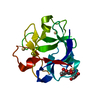
| ||||||||
|---|---|---|---|---|---|---|---|---|---|
| 1 |
| ||||||||
| Unit cell |
| ||||||||
| Components on special symmetry positions |
|
-Components
| #1: Protein | Mass: 13933.389 Da / Num. of mol.: 1 Source method: isolated from a genetically manipulated source Details: Residue 6-132 of the mature protein preceded by residues left from TEV cleavage site. We used the numbering such as we kept the one of the mature protein Source: (gene. exp.) Cucumis melo (muskmelon) / Gene: LOC107992255, 107992255 / Production host:  | ||||||
|---|---|---|---|---|---|---|---|
| #2: Polysaccharide | beta-D-galactopyranose-(1-4)-2-acetamido-2-deoxy-alpha-D-glucopyranose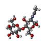  Source method: isolated from a genetically manipulated source Details: oligosaccharide / References: N-acetyl-alpha-lactosamine | ||||||
| #3: Chemical |   Mass: 112.411 Da / Num. of mol.: 3 / Source method: obtained synthetically / Formula: Cd / Feature type: SUBJECT OF INVESTIGATION Mass: 112.411 Da / Num. of mol.: 3 / Source method: obtained synthetically / Formula: Cd / Feature type: SUBJECT OF INVESTIGATION#4: Water | ChemComp-HOH / |  Mass: 18.015 Da / Num. of mol.: 241 / Source method: isolated from a natural source / Formula: H2O Mass: 18.015 Da / Num. of mol.: 241 / Source method: isolated from a natural source / Formula: H2OHas ligand of interest | Y | Has protein modification | Y | |
-Experimental details
-Experiment
| Experiment | Method: X-RAY DIFFRACTION / Number of used crystals: 1 |
|---|
- Sample preparation
| Crystal | Density Matthews: 2.29 Å3/Da / Density % sol: 46.21 % / Description: diamond |
|---|---|
| Crystal grow | Temperature: 292 K / Method: vapor diffusion, hanging drop Details: 10% Peg Smear Medium, 0.1 M Mes pH 6.5, and 5 mM of CaCl2, MgCl2, CsCl2, CdCl2, NiCl2 and Zinc acetate transfered in 30% Peg Smear Medium 5mM CdCl2 for cryoprotection |
-Data collection
| Diffraction | Mean temperature: 100 K / Serial crystal experiment: N |
|---|---|
| Diffraction source | Source: SYNCHROTRON / Site: SOLEIL  / Beamline: PROXIMA 1 / Wavelength: 0.97856 Å / Beamline: PROXIMA 1 / Wavelength: 0.97856 Å |
| Detector | Type: DECTRIS EIGER X 16M / Detector: PIXEL / Date: Oct 5, 2023 |
| Radiation | Protocol: SINGLE WAVELENGTH / Monochromatic (M) / Laue (L): M / Scattering type: x-ray |
| Radiation wavelength | Wavelength: 0.97856 Å / Relative weight: 1 |
| Reflection | Resolution: 1.317→46.78 Å / Num. obs: 29695 / % possible obs: 99.8 % / Redundancy: 13.5 % / CC1/2: 0.999 / Rmerge(I) obs: 0.055 / Rpim(I) all: 0.022 / Rrim(I) all: 0.059 / Net I/σ(I): 25.2 |
| Reflection shell | Resolution: 1.32→1.34 Å / Rmerge(I) obs: 0.369 / Mean I/σ(I) obs: 5.7 / Num. unique obs: 1434 / CC1/2: 0.969 / Rpim(I) all: 0.153 / Rrim(I) all: 0.4 |
- Processing
| Software |
| |||||||||||||||||||||||||||||||||||||||||||||||||||||||||||||||||||||||||||||||||||||||||||||||||||||||||||||||||||||||||||||||||||||||||||||||||||||||||||||||||||||||||||||||||||||||||||||||||||||||||||||||||||||||||||||||||||||||
|---|---|---|---|---|---|---|---|---|---|---|---|---|---|---|---|---|---|---|---|---|---|---|---|---|---|---|---|---|---|---|---|---|---|---|---|---|---|---|---|---|---|---|---|---|---|---|---|---|---|---|---|---|---|---|---|---|---|---|---|---|---|---|---|---|---|---|---|---|---|---|---|---|---|---|---|---|---|---|---|---|---|---|---|---|---|---|---|---|---|---|---|---|---|---|---|---|---|---|---|---|---|---|---|---|---|---|---|---|---|---|---|---|---|---|---|---|---|---|---|---|---|---|---|---|---|---|---|---|---|---|---|---|---|---|---|---|---|---|---|---|---|---|---|---|---|---|---|---|---|---|---|---|---|---|---|---|---|---|---|---|---|---|---|---|---|---|---|---|---|---|---|---|---|---|---|---|---|---|---|---|---|---|---|---|---|---|---|---|---|---|---|---|---|---|---|---|---|---|---|---|---|---|---|---|---|---|---|---|---|---|---|---|---|---|---|---|---|---|---|---|---|---|---|---|---|---|---|---|---|---|---|---|
| Refinement | Method to determine structure: MOLECULAR REPLACEMENT / Resolution: 1.317→46.778 Å / Cor.coef. Fo:Fc: 0.971 / Cor.coef. Fo:Fc free: 0.95 / SU B: 1.297 / SU ML: 0.025 / Cross valid method: FREE R-VALUE / ESU R: 0.054 / ESU R Free: 0.054 Details: Hydrogens have been added in their riding positions anisotropic refinement
| |||||||||||||||||||||||||||||||||||||||||||||||||||||||||||||||||||||||||||||||||||||||||||||||||||||||||||||||||||||||||||||||||||||||||||||||||||||||||||||||||||||||||||||||||||||||||||||||||||||||||||||||||||||||||||||||||||||||
| Solvent computation | Ion probe radii: 0.8 Å / Shrinkage radii: 0.8 Å / VDW probe radii: 1.2 Å / Solvent model: MASK BULK SOLVENT | |||||||||||||||||||||||||||||||||||||||||||||||||||||||||||||||||||||||||||||||||||||||||||||||||||||||||||||||||||||||||||||||||||||||||||||||||||||||||||||||||||||||||||||||||||||||||||||||||||||||||||||||||||||||||||||||||||||||
| Displacement parameters | Biso mean: 13.471 Å2
| |||||||||||||||||||||||||||||||||||||||||||||||||||||||||||||||||||||||||||||||||||||||||||||||||||||||||||||||||||||||||||||||||||||||||||||||||||||||||||||||||||||||||||||||||||||||||||||||||||||||||||||||||||||||||||||||||||||||
| Refinement step | Cycle: LAST / Resolution: 1.317→46.778 Å
| |||||||||||||||||||||||||||||||||||||||||||||||||||||||||||||||||||||||||||||||||||||||||||||||||||||||||||||||||||||||||||||||||||||||||||||||||||||||||||||||||||||||||||||||||||||||||||||||||||||||||||||||||||||||||||||||||||||||
| Refine LS restraints |
| |||||||||||||||||||||||||||||||||||||||||||||||||||||||||||||||||||||||||||||||||||||||||||||||||||||||||||||||||||||||||||||||||||||||||||||||||||||||||||||||||||||||||||||||||||||||||||||||||||||||||||||||||||||||||||||||||||||||
| LS refinement shell | Refine-ID: X-RAY DIFFRACTION / Total num. of bins used: 20
|
