[English] 日本語
Yorodumi
- PDB-8r5w: Crystal structure of the three anaphylatoxin-like modules in fibulin-2 -

+
Open data


ID or keywords:

Loading...

-
Basic information

Entry
Database: PDB / ID: 8r5w
TitleCrystal structure of the three anaphylatoxin-like modules in fibulin-2
ComponentsFibulin-2
KeywordsSTRUCTURAL PROTEIN / Extracellular matrix / Fibulins / anaphylatoxin-like
Function / homology
Function and homology information


Molecules associated with elastic fibres / extracellular matrix binding / positive regulation of cell-substrate adhesion / collagen-containing extracellular matrix / calcium ion binding / extracellular region
Similarity search - Function
Complement Clr-like EGF domain / Complement Clr-like EGF-like / Anaphylatoxin domain signature. / Anaphylatoxin/fibulin / Anaphylotoxin-like domain / Anaphylatoxin domain profile. / Anaphylatoxin homologous domain / : / Calcium-binding EGF domain / Coagulation Factor Xa inhibitory site ...Complement Clr-like EGF domain / Complement Clr-like EGF-like / Anaphylatoxin domain signature. / Anaphylatoxin/fibulin / Anaphylotoxin-like domain / Anaphylatoxin domain profile. / Anaphylatoxin homologous domain / : / Calcium-binding EGF domain / Coagulation Factor Xa inhibitory site / EGF-type aspartate/asparagine hydroxylation site / EGF-like calcium-binding, conserved site / Calcium-binding EGF-like domain signature. / Aspartic acid and asparagine hydroxylation site. / EGF-like calcium-binding domain / Calcium-binding EGF-like domain / Epidermal growth factor-like domain. / EGF-like domain profile. / Growth factor receptor cysteine-rich domain superfamily / EGF-like domain signature 2. / EGF-like domain
Similarity search - Domain/homology
Biological speciesMus musculus (house mouse)
MethodX-RAY DIFFRACTION / SYNCHROTRON / MOLECULAR REPLACEMENT / Resolution: 2.2 Å
AuthorsSohail, A.A. / Koski, M.K. / Ruddock, L.W.
Funding support Finland, 1items
OrganizationGrant numberCountry
Academy of Finland272573 Finland
CitationJournal: Sci Rep / Year: 2024
Title: Biophysical and structural studies of fibulin-2
Authors: Sohail, A.A. / Koski, M.K. / Ruddock, L.W.
History
DepositionNov 18, 2023Deposition site: PDBE / Processing site: PDBE
Revision 1.0Jul 10, 2024Provider: repository / Type: Initial release

-
Structure visualization

Structure viewerMolecule:
MolmilJmol/JSmol

Downloads & links

-
Assembly

Deposited unit
A: Fibulin-2
B: Fibulin-2
C: Fibulin-2
D: Fibulin-2
E: Fibulin-2
F: Fibulin-2
G: Fibulin-2
H: Fibulin-2


Theoretical massNumber of molelcules
Total (without water)110,6618
Polymers110,6618
Non-polymers00
Water1,946108
1


  • Idetical with deposited unit
  • defined by author&software
  • Evidence: light scattering, Static light scattering was done to show that the molecule forms a dimer.
TypeNameSymmetry operationNumber
identity operation1_555x,y,z1
Unit cell
Length a, b, c (Å)103.680, 34.510, 106.890
Angle α, β, γ (deg.)90.00, 94.00, 90.00
Int Tables number4
Space group name H-MP1211

-
Components

#1: Protein
Fibulin-2


Mass: 13832.585 Da / Num. of mol.: 8
Source method: isolated from a genetically manipulated source
Source: (gene. exp.) Mus musculus (house mouse) / Gene: Fbln2 / Production host: Escherichia coli B (bacteria) / References: UniProt: P37889
#2: Water ChemComp-HOH / water


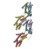
Mass: 18.015 Da / Num. of mol.: 108 / Source method: isolated from a natural source / Formula: H2O

-
Experimental details

-
Experiment

ExperimentMethod: X-RAY DIFFRACTION / Number of used crystals: 1

-
Sample preparation

CrystalDensity Matthews: 1.72 Å3/Da / Density % sol: 28.65 %
Crystal growTemperature: 295 K / Method: vapor diffusion, sitting drop / pH: 8 / Details: 0.2 M potassium nitrate, 2 % w/v PEG 3350

-
Data collection

DiffractionMean temperature: 100 K / Serial crystal experiment: N
Diffraction sourceSource: SYNCHROTRON / Site: Diamond / Beamline: I04 / Wavelength: 0.9537 Å
DetectorType: DECTRIS EIGER2 XE 16M / Detector: PIXEL / Date: Jan 20, 2023
RadiationProtocol: SINGLE WAVELENGTH / Monochromatic (M) / Laue (L): M / Scattering type: x-ray
Radiation wavelengthWavelength: 0.9537 Å / Relative weight: 1
ReflectionResolution: 1.97→48.8 Å / Num. obs: 54442 / % possible obs: 99.87 % / Redundancy: 6.8 % / CC1/2: 0.9993 / Net I/σ(I): 8.4
Reflection shellResolution: 1.97→2 Å / Redundancy: 7.1 % / Mean I/σ(I) obs: 0.7 / Num. unique obs: 2700 / CC1/2: 0.329 / % possible all: 99.93

-
Processing

Software
NameVersionClassification
REFMAC5.8.0419refinement
xia2data reduction
xia2data scaling
PHENIXphasing
RefinementMethod to determine structure: MOLECULAR REPLACEMENT / Resolution: 2.2→48.8 Å / Cor.coef. Fo:Fc: 0.946 / Cor.coef. Fo:Fc free: 0.911 / SU B: 20.487 / SU ML: 0.236 / Cross valid method: THROUGHOUT / ESU R: 0.424 / ESU R Free: 0.28 / Stereochemistry target values: MAXIMUM LIKELIHOOD / Details: HYDROGENS HAVE BEEN USED IF PRESENT IN THE INPUT
RfactorNum. reflection% reflectionSelection details
Rfree0.29742 1998 5.1 %RANDOM
Rwork0.24279 ---
obs0.24544 37274 99.8 %-
Solvent computationIon probe radii: 0.8 Å / Shrinkage radii: 0.8 Å / VDW probe radii: 1.2 Å / Solvent model: MASK
Displacement parametersBiso mean: 57.605 Å2
Baniso -1Baniso -2Baniso -3
1-2.79 Å20 Å20.47 Å2
2--0.9 Å2-0 Å2
3----3.72 Å2
Refinement stepCycle: 1 / Resolution: 2.2→48.8 Å
ProteinNucleic acidLigandSolventTotal
Num. atoms6449 0 0 108 6557
Refine LS restraints
Refine-IDTypeDev idealDev ideal targetNumber
X-RAY DIFFRACTIONr_bond_refined_d0.0080.0126624
X-RAY DIFFRACTIONr_bond_other_d0.0010.0165966
X-RAY DIFFRACTIONr_angle_refined_deg1.5321.8178921
X-RAY DIFFRACTIONr_angle_other_deg0.5291.7813869
X-RAY DIFFRACTIONr_dihedral_angle_1_deg5.135861
X-RAY DIFFRACTIONr_dihedral_angle_2_deg7.217535
X-RAY DIFFRACTIONr_dihedral_angle_3_deg16.696101187
X-RAY DIFFRACTIONr_dihedral_angle_4_deg
X-RAY DIFFRACTIONr_chiral_restr0.0820.2958
X-RAY DIFFRACTIONr_gen_planes_refined0.0060.027862
X-RAY DIFFRACTIONr_gen_planes_other0.0010.021386
X-RAY DIFFRACTIONr_nbd_refined
X-RAY DIFFRACTIONr_nbd_other
X-RAY DIFFRACTIONr_nbtor_refined
X-RAY DIFFRACTIONr_nbtor_other
X-RAY DIFFRACTIONr_xyhbond_nbd_refined
X-RAY DIFFRACTIONr_xyhbond_nbd_other
X-RAY DIFFRACTIONr_metal_ion_refined
X-RAY DIFFRACTIONr_metal_ion_other
X-RAY DIFFRACTIONr_symmetry_vdw_refined
X-RAY DIFFRACTIONr_symmetry_vdw_other
X-RAY DIFFRACTIONr_symmetry_hbond_refined
X-RAY DIFFRACTIONr_symmetry_hbond_other
X-RAY DIFFRACTIONr_symmetry_metal_ion_refined
X-RAY DIFFRACTIONr_symmetry_metal_ion_other
X-RAY DIFFRACTIONr_mcbond_it4.1434.0033474
X-RAY DIFFRACTIONr_mcbond_other4.1434.0033474
X-RAY DIFFRACTIONr_mcangle_it6.1147.1414304
X-RAY DIFFRACTIONr_mcangle_other6.1147.1434305
X-RAY DIFFRACTIONr_scbond_it5.1664.7283150
X-RAY DIFFRACTIONr_scbond_other5.1624.7273149
X-RAY DIFFRACTIONr_scangle_it
X-RAY DIFFRACTIONr_scangle_other8.0268.4434611
X-RAY DIFFRACTIONr_long_range_B_refined10.69250.587452
X-RAY DIFFRACTIONr_long_range_B_other10.69550.497443
X-RAY DIFFRACTIONr_rigid_bond_restr
X-RAY DIFFRACTIONr_sphericity_free
X-RAY DIFFRACTIONr_sphericity_bonded
LS refinement shellResolution: 2.2→2.257 Å / Total num. of bins used: 20
RfactorNum. reflection% reflection
Rfree0.329 120 -
Rwork0.329 2740 -
obs--100 %
Refinement TLS params.

Method: refined / Refine-ID: X-RAY DIFFRACTION

IDL112)L122)L132)L222)L232)L332)S11 (Å °)S12 (Å °)S13 (Å °)S21 (Å °)S22 (Å °)S23 (Å °)S31 (Å °)S32 (Å °)S33 (Å °)T112)T122)T132)T222)T232)T332)Origin x (Å)Origin y (Å)Origin z (Å)
11.8222-0.56481.17650.8234-0.49931.5614-0.0227-0.06580.04870.03710.0250.0993-0.0902-0.1186-0.00230.1028-0.02160.01090.08350.01080.172840.22230.526718.9084
22.3118-1.10621.26451.1515-1.12013.13220.00010.01190.0591-0.07910.06540.0122-0.0531-0.0308-0.06550.0611-0.0162-0.01670.0716-0.02810.09549.66371.54169.4122
32.23411.45131.23892.42921.86893.02670.04890.00020.0176-0.0249-0.0025-0.0893-0.08810.005-0.04640.01030.01040.01660.10990.01230.065175.673416.684944.3645
41.43531.10960.96191.76621.35412.0917-0.01050.035-0.07-0.12510.0275-0.201-0.07360.0062-0.0170.08590.03440.0630.13240.01010.210486.278616.16936.1116
51.5512-0.56221.21770.7599-0.79471.5886-0.0763-0.07820.00150.06170.08030.053-0.1278-0.1494-0.00410.1417-0.01820.03390.1653-0.04930.170562.10120.114272.9264
62.3746-0.81720.88351.5139-1.07562.3377-0.01660.01270.0978-0.01390.0234-0.1058-0.05470.0282-0.00680.0429-0.0226-0.00040.1254-0.07550.062471.37550.403363.0876
72.31751.01230.95231.69881.51663.02110.00240.0609-0.0099-0.03650.0478-0.0122-0.1302-0.0206-0.05020.01540.0177-0.00010.04760.03720.09198.333616.043797.3747
81.54410.83590.3691.64671.08392.334-0.08770.0230.0334-0.05840.0322-0.0011-0.0220.23850.05540.09370.03720.04470.11580.00930.1582108.99216.342188.9117
Refinement TLS group
IDRefine-IDRefine TLS-IDAuth asym-IDAuth seq-ID
1X-RAY DIFFRACTION1A419 - 545
2X-RAY DIFFRACTION2B427 - 545
3X-RAY DIFFRACTION3C427 - 545
4X-RAY DIFFRACTION4D423 - 544
5X-RAY DIFFRACTION5E419 - 544
6X-RAY DIFFRACTION6F427 - 545
7X-RAY DIFFRACTION7G427 - 545
8X-RAY DIFFRACTION8H426 - 544

+
About Yorodumi

-
News

-
Feb 9, 2022. New format data for meta-information of EMDB entries

New format data for meta-information of EMDB entries

  • Version 3 of the EMDB header file is now the official format.
  • The previous official version 1.9 will be removed from the archive.

Related info.:EMDB header

External links:wwPDB to switch to version 3 of the EMDB data model

-
Aug 12, 2020. Covid-19 info

Covid-19 info

URL: https://pdbjlvh1.pdbj.org/emnavi/covid19.php

New page: Covid-19 featured information page in EM Navigator.

Related info.:Covid-19 info / Mar 5, 2020. Novel coronavirus structure data

+
Mar 5, 2020. Novel coronavirus structure data

Novel coronavirus structure data

Related info.:Yorodumi Speices / Aug 12, 2020. Covid-19 info

External links:COVID-19 featured content - PDBj / Molecule of the Month (242):Coronavirus Proteases

+
Jan 31, 2019. EMDB accession codes are about to change! (news from PDBe EMDB page)

EMDB accession codes are about to change! (news from PDBe EMDB page)

  • The allocation of 4 digits for EMDB accession codes will soon come to an end. Whilst these codes will remain in use, new EMDB accession codes will include an additional digit and will expand incrementally as the available range of codes is exhausted. The current 4-digit format prefixed with “EMD-” (i.e. EMD-XXXX) will advance to a 5-digit format (i.e. EMD-XXXXX), and so on. It is currently estimated that the 4-digit codes will be depleted around Spring 2019, at which point the 5-digit format will come into force.
  • The EM Navigator/Yorodumi systems omit the EMD- prefix.

Related info.:Q: What is EMD? / ID/Accession-code notation in Yorodumi/EM Navigator

External links:EMDB Accession Codes are Changing Soon! / Contact to PDBj

+
Jul 12, 2017. Major update of PDB

Major update of PDB

  • wwPDB released updated PDB data conforming to the new PDBx/mmCIF dictionary.
  • This is a major update changing the version number from 4 to 5, and with Remediation, in which all the entries are updated.
  • In this update, many items about electron microscopy experimental information are reorganized (e.g. em_software).
  • Now, EM Navigator and Yorodumi are based on the updated data.

External links:wwPDB Remediation / Enriched Model Files Conforming to OneDep Data Standards Now Available in the PDB FTP Archive

-
Yorodumi

Thousand views of thousand structures

  • Yorodumi is a browser for structure data from EMDB, PDB, SASBDB, etc.
  • This page is also the successor to EM Navigator detail page, and also detail information page/front-end page for Omokage search.
  • The word "yorodu" (or yorozu) is an old Japanese word meaning "ten thousand". "mi" (miru) is to see.

Related info.:EMDB / PDB / SASBDB / Comparison of 3 databanks / Yorodumi Search / Aug 31, 2016. New EM Navigator & Yorodumi / Yorodumi Papers / Jmol/JSmol / Function and homology information / Changes in new EM Navigator and Yorodumi

Read more