 Movie
Movie Controller

[English] 日本語
 Yorodumi
- PDB-8r49: Plastidial phosphorylase Pho1 from Solanum tuberosum in complex w... -
+ Open data

- Basic information
| Entry | Database: PDB / ID: 8r49 | ||||||
|---|---|---|---|---|---|---|---|
| Title | Plastidial phosphorylase Pho1 from Solanum tuberosum in complex with beta cyclodextrin | ||||||
 Components Components | Alpha-1,4 glucan phosphorylase L-1 isozyme, chloroplastic/amyloplastic | ||||||
 Keywords Keywords | TRANSFERASE / starch phosphorylase / plastidial phosphorylase / carbohydrate metabolsim | ||||||
| Function / homology |  Function and homology information Function and homology informationamyloplast / glycogen phosphorylase / glycogen phosphorylase activity / glycogen catabolic process / chloroplast / pyridoxal phosphate binding / identical protein binding / cytoplasm Similarity search - Function | ||||||
| Biological species |  | ||||||
| Method |  X-RAY DIFFRACTION / SYNCHROTRON / MOLECULAR REPLACEMENT / Resolution: 2.8 Å X-RAY DIFFRACTION / SYNCHROTRON / MOLECULAR REPLACEMENT / Resolution: 2.8 Å | ||||||
 Authors Authors | Koulas, S.M. / Leonidas, D.D. | ||||||
| Funding support |  Greece, 1items Greece, 1items
| ||||||
 Citation Citation | Journal: Acs Omega / Year: 2024 Title: Kinetic and Structural Studies of the Plastidial Solanum tuberosum Phosphorylase. Authors: Koulas, S.M. / Kyriakis, E. / Tsagkarakou, A.S. / Leonidas, D.D. | ||||||
| History |
|
- Structure visualization
| Structure viewer | Molecule: MolmilJmol/JSmol |
|---|
- Downloads & links
-Download
| PDBx/mmCIF format | 8r49.cif.gz | 528.6 KB | Display | PDBx/mmCIF format |
|---|---|---|---|---|
| PDB format | pdb8r49.ent.gz | 413.2 KB | Display | PDB format |
| PDBx/mmJSON format | 8r49.json.gz | Tree view | PDBx/mmJSON format | |
| Others |  Other downloads Other downloads |
-Validation report
| Arichive directory | https://data.pdbj.org/pub/pdb/validation_reports/r4/8r49ftp://data.pdbj.org/pub/pdb/validation_reports/r4/8r49 | HTTPS FTP |
|---|
-Related structure data


-Links
 PDBj
PDBj

- Assembly
| Deposited unit | 
| ||||||||||||||||||||||||||||||||||||||||||||||||||
|---|---|---|---|---|---|---|---|---|---|---|---|---|---|---|---|---|---|---|---|---|---|---|---|---|---|---|---|---|---|---|---|---|---|---|---|---|---|---|---|---|---|---|---|---|---|---|---|---|---|---|---|
| 1 | 
| ||||||||||||||||||||||||||||||||||||||||||||||||||
| 2 | 
| ||||||||||||||||||||||||||||||||||||||||||||||||||
| Unit cell |
| ||||||||||||||||||||||||||||||||||||||||||||||||||
| Noncrystallographic symmetry (NCS) | NCS domain:
NCS domain segments: Beg auth comp-ID: ALA / Beg label comp-ID: ALA / End auth comp-ID: ALA / End label comp-ID: ALA / Auth asym-ID: A / Label asym-ID: A / Auth seq-ID: 23 - 916 / Label seq-ID: 23 - 916
NCS ensembles :
|
-Components
| #1: Protein | Mass: 104008.430 Da / Num. of mol.: 3 / Source method: isolated from a natural source / Source: (natural) #2: Polysaccharide | 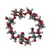  Type: oligosaccharide, Oligosaccharide / Class: Drug delivery / Mass: 1153.001 Da / Num. of mol.: 3 Type: oligosaccharide, Oligosaccharide / Class: Drug delivery / Mass: 1153.001 Da / Num. of mol.: 3Source method: isolated from a genetically manipulated source Details: cyclic oligosaccharide / References: beta-cyclodextrin #3: Chemical | ChemComp-DMS /   Mass: 78.133 Da / Num. of mol.: 8 Mass: 78.133 Da / Num. of mol.: 8Source method: isolated from a genetically manipulated source Formula: C2H6OS / Comment: DMSO, precipitant*YM #4: Water | ChemComp-HOH / |  Mass: 18.015 Da / Num. of mol.: 101 / Source method: isolated from a natural source / Formula: H2O Mass: 18.015 Da / Num. of mol.: 101 / Source method: isolated from a natural source / Formula: H2OHas ligand of interest | Y | Has protein modification | Y | |
|---|
-Experimental details
-Experiment
| Experiment | Method: X-RAY DIFFRACTION / Number of used crystals: 1 |
|---|
- Sample preparation
| Crystal | Density Matthews: 2.96 Å3/Da / Density % sol: 58.38 % |
|---|---|
| Crystal grow | Temperature: 289 K / Method: vapor diffusion, sitting drop / pH: 8.5 / Details: 0.1 M Tris-HCl, 20 % PEG4000 |
-Data collection
| Diffraction | Mean temperature: 100 K / Serial crystal experiment: N |
|---|---|
| Diffraction source | Source: SYNCHROTRON / Site: PETRA III, EMBL c/o DESY  / Beamline: P13 (MX1) / Wavelength: 0.976256 Å / Beamline: P13 (MX1) / Wavelength: 0.976256 Å |
| Detector | Type: DECTRIS PILATUS 6M / Detector: PIXEL / Date: Jun 26, 2023 |
| Radiation | Protocol: SINGLE WAVELENGTH / Monochromatic (M) / Laue (L): M / Scattering type: x-ray |
| Radiation wavelength | Wavelength: 0.976256 Å / Relative weight: 1 |
| Reflection | Resolution: 2.8→123.114 Å / Num. obs: 87204 / % possible obs: 98 % / Redundancy: 3 % / CC1/2: 0.995 / Rmerge(I) obs: 0.09 / Net I/σ(I): 7.4 |
| Reflection shell | Resolution: 2.8→2.85 Å / Rmerge(I) obs: 0.818 / Num. unique obs: 4507 / CC1/2: 0.654 |
- Processing
| Software |
| |||||||||||||||||||||||||||||||||||||||||||||||||||||||||||||||||||||||||||||||||||||||||||||||||||||||||||||||||||||||||||||||||||||||||||||||||||||||||||||||||||||||||||||||||||||||||||||||||||||||||||||||||||||||||||||||||||||||
|---|---|---|---|---|---|---|---|---|---|---|---|---|---|---|---|---|---|---|---|---|---|---|---|---|---|---|---|---|---|---|---|---|---|---|---|---|---|---|---|---|---|---|---|---|---|---|---|---|---|---|---|---|---|---|---|---|---|---|---|---|---|---|---|---|---|---|---|---|---|---|---|---|---|---|---|---|---|---|---|---|---|---|---|---|---|---|---|---|---|---|---|---|---|---|---|---|---|---|---|---|---|---|---|---|---|---|---|---|---|---|---|---|---|---|---|---|---|---|---|---|---|---|---|---|---|---|---|---|---|---|---|---|---|---|---|---|---|---|---|---|---|---|---|---|---|---|---|---|---|---|---|---|---|---|---|---|---|---|---|---|---|---|---|---|---|---|---|---|---|---|---|---|---|---|---|---|---|---|---|---|---|---|---|---|---|---|---|---|---|---|---|---|---|---|---|---|---|---|---|---|---|---|---|---|---|---|---|---|---|---|---|---|---|---|---|---|---|---|---|---|---|---|---|---|---|---|---|---|---|---|---|---|
| Refinement | Method to determine structure: MOLECULAR REPLACEMENT / Resolution: 2.8→123.114 Å / Cor.coef. Fo:Fc: 0.949 / Cor.coef. Fo:Fc free: 0.942 / WRfactor Rfree: 0.239 / WRfactor Rwork: 0.224 / SU B: 19.3 / SU ML: 0.332 / Average fsc free: 0.9443 / Average fsc work: 0.951 / Cross valid method: THROUGHOUT / ESU R: 2.486 / ESU R Free: 0.34 Details: Hydrogens have been added in their riding positions
| |||||||||||||||||||||||||||||||||||||||||||||||||||||||||||||||||||||||||||||||||||||||||||||||||||||||||||||||||||||||||||||||||||||||||||||||||||||||||||||||||||||||||||||||||||||||||||||||||||||||||||||||||||||||||||||||||||||||
| Solvent computation | Ion probe radii: 0.9 Å / Shrinkage radii: 0.9 Å / VDW probe radii: 1 Å / Solvent model: MASK BULK SOLVENT | |||||||||||||||||||||||||||||||||||||||||||||||||||||||||||||||||||||||||||||||||||||||||||||||||||||||||||||||||||||||||||||||||||||||||||||||||||||||||||||||||||||||||||||||||||||||||||||||||||||||||||||||||||||||||||||||||||||||
| Displacement parameters | Biso mean: 88.194 Å2
| |||||||||||||||||||||||||||||||||||||||||||||||||||||||||||||||||||||||||||||||||||||||||||||||||||||||||||||||||||||||||||||||||||||||||||||||||||||||||||||||||||||||||||||||||||||||||||||||||||||||||||||||||||||||||||||||||||||||
| Refinement step | Cycle: LAST / Resolution: 2.8→123.114 Å
| |||||||||||||||||||||||||||||||||||||||||||||||||||||||||||||||||||||||||||||||||||||||||||||||||||||||||||||||||||||||||||||||||||||||||||||||||||||||||||||||||||||||||||||||||||||||||||||||||||||||||||||||||||||||||||||||||||||||
| Refine LS restraints |
| |||||||||||||||||||||||||||||||||||||||||||||||||||||||||||||||||||||||||||||||||||||||||||||||||||||||||||||||||||||||||||||||||||||||||||||||||||||||||||||||||||||||||||||||||||||||||||||||||||||||||||||||||||||||||||||||||||||||
| Refine LS restraints NCS |
| |||||||||||||||||||||||||||||||||||||||||||||||||||||||||||||||||||||||||||||||||||||||||||||||||||||||||||||||||||||||||||||||||||||||||||||||||||||||||||||||||||||||||||||||||||||||||||||||||||||||||||||||||||||||||||||||||||||||
| LS refinement shell | Refine-ID: X-RAY DIFFRACTION / Total num. of bins used: 20
|
