 Movie
Movie Controller


+ Open data

- Basic information
| Entry | Database: PDB / ID: 8r15 | ||||||
|---|---|---|---|---|---|---|---|
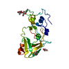
| Title | Crystal structure of Fusarium oxysporum NADase I | ||||||
 Components Components | TNT domain-containing protein | ||||||
 Keywords Keywords | HYDROLASE / NADase / NAD hydrolase / homodimer / glycoprotein / extracellular / TNT domain / covalent dimer | ||||||
| Function / homology | : / Tuberculosis necrotizing toxin / Tuberculosis necrotizing toxin / NADP+ nucleosidase activity / TNT domain-containing protein Function and homology information Function and homology information | ||||||
| Biological species |  Fusarium oxysporum f. sp. cubense race 1 (fungus) Fusarium oxysporum f. sp. cubense race 1 (fungus) | ||||||
| Method |  X-RAY DIFFRACTION / SYNCHROTRON / MOLECULAR REPLACEMENT / Resolution: 2.4 Å X-RAY DIFFRACTION / SYNCHROTRON / MOLECULAR REPLACEMENT / Resolution: 2.4 Å | ||||||
 Authors Authors | Kallio, J.P. / Ferrario, E. / Stromland, O. / Ziegler, M. | ||||||
| Funding support |  Norway, 1items Norway, 1items
| ||||||
 Citation Citation | Journal: Protein Sci. / Year: 2024 Title: Evolution of fungal tuberculosis necrotizing toxin (TNT) domain-containing enzymes reveals divergent adaptations to enhance NAD cleavage. Authors: Ferrario, E. / Kallio, J.P. / Emdadi, M. / Stromland, O. / Rack, J.G.M. / Ziegler, M. #1: Journal: Acta Crystallogr.,Sect.D / Year: 2012 Title: Towards automated crystallographic structure refinement with phenix.refine. Authors: Afonine, P.V. / Grosse-Kunstleve, R.W. / Echols, N. / Headd, J.J. / Moriarty, N.W. / Mustyakimov, M. / Terwilliger, T.C. / Urzhumtsev, A. / Zwart, P.H. / Adams, P.D. #2: Journal: Acta Crystallogr D Struct Biol / Year: 2019 Title: Macromolecular structure determination using X-rays, neutrons and electrons: recent developments in Phenix. Authors: Dorothee Liebschner / Pavel V Afonine / Matthew L Baker / Gábor Bunkóczi / Vincent B Chen / Tristan I Croll / Bradley Hintze / Li Wei Hung / Swati Jain / Airlie J McCoy / Nigel W Moriarty ...Authors: Dorothee Liebschner / Pavel V Afonine / Matthew L Baker / Gábor Bunkóczi / Vincent B Chen / Tristan I Croll / Bradley Hintze / Li Wei Hung / Swati Jain / Airlie J McCoy / Nigel W Moriarty / Robert D Oeffner / Billy K Poon / Michael G Prisant / Randy J Read / Jane S Richardson / David C Richardson / Massimo D Sammito / Oleg V Sobolev / Duncan H Stockwell / Thomas C Terwilliger / Alexandre G Urzhumtsev / Lizbeth L Videau / Christopher J Williams / Paul D Adams /    Abstract: Diffraction (X-ray, neutron and electron) and electron cryo-microscopy are powerful methods to determine three-dimensional macromolecular structures, which are required to understand biological ...Diffraction (X-ray, neutron and electron) and electron cryo-microscopy are powerful methods to determine three-dimensional macromolecular structures, which are required to understand biological processes and to develop new therapeutics against diseases. The overall structure-solution workflow is similar for these techniques, but nuances exist because the properties of the reduced experimental data are different. Software tools for structure determination should therefore be tailored for each method. Phenix is a comprehensive software package for macromolecular structure determination that handles data from any of these techniques. Tasks performed with Phenix include data-quality assessment, map improvement, model building, the validation/rebuilding/refinement cycle and deposition. Each tool caters to the type of experimental data. The design of Phenix emphasizes the automation of procedures, where possible, to minimize repetitive and time-consuming manual tasks, while default parameters are chosen to encourage best practice. A graphical user interface provides access to many command-line features of Phenix and streamlines the transition between programs, project tracking and re-running of previous tasks. | ||||||
| History |
|
- Structure visualization
| Structure viewer | Molecule: MolmilJmol/JSmol |
|---|
- Downloads & links
-Download
| PDBx/mmCIF format | 8r15.cif.gz | 115.4 KB | Display | PDBx/mmCIF format |
|---|---|---|---|---|
| PDB format | pdb8r15.ent.gz | 72.9 KB | Display | PDB format |
| PDBx/mmJSON format | 8r15.json.gz | Tree view | PDBx/mmJSON format | |
| Others |  Other downloads Other downloads |
-Validation report
| Summary document | 8r15_validation.pdf.gz | 769.8 KB | Display | wwPDB validaton report |
|---|---|---|---|---|
| Full document | 8r15_full_validation.pdf.gz | 771 KB | Display | |
| Data in XML | 8r15_validation.xml.gz | 10.6 KB | Display | |
| Data in CIF | 8r15_validation.cif.gz | 13.8 KB | Display | |
| Arichive directory | https://data.pdbj.org/pub/pdb/validation_reports/r1/8r15ftp://data.pdbj.org/pub/pdb/validation_reports/r1/8r15 | HTTPS FTP |
-Related structure data
-Links
 PDBj
PDBj- Assembly
| Deposited unit | 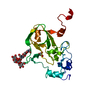
| ||||||||||||
|---|---|---|---|---|---|---|---|---|---|---|---|---|---|
| 1 | 
| ||||||||||||
| Unit cell |
| ||||||||||||
| Components on special symmetry positions |
|
-Components
| #1: Protein | Mass: 24824.961 Da / Num. of mol.: 1 Source method: isolated from a genetically manipulated source Details: MLPSFLLSSLLCLSTVSA = signal sequence DVLFQGPGHHHHHH= 3c protease site and His-tag Source: (gene. exp.) Fusarium oxysporum f. sp. cubense race 1 (fungus)Gene: FOC1_g10001035 / Production host:   Spodoptera frugiperda (fall armyworm) / References: UniProt: N4U1S7 Spodoptera frugiperda (fall armyworm) / References: UniProt: N4U1S7 |
|---|---|
| #2: Polysaccharide | alpha-D-mannopyranose-(1-3)-[alpha-D-mannopyranose-(1-6)]beta-D-mannopyranose-(1-4)-2-acetamido-2- ...alpha-D-mannopyranose-(1-3)-[alpha-D-mannopyranose-(1-6)]beta-D-mannopyranose-(1-4)-2-acetamido-2-deoxy-beta-D-glucopyranose-(1-4)-2-acetamido-2-deoxy-beta-D-glucopyranose Source method: isolated from a genetically manipulated source |
| #3: Chemical | ChemComp-GOL /   Mass: 92.094 Da / Num. of mol.: 1 / Source method: obtained synthetically / Formula: C3H8O3 Mass: 92.094 Da / Num. of mol.: 1 / Source method: obtained synthetically / Formula: C3H8O3 |
| #4: Water | ChemComp-HOH /  Mass: 18.015 Da / Num. of mol.: 70 / Source method: isolated from a natural source / Formula: H2O Mass: 18.015 Da / Num. of mol.: 70 / Source method: isolated from a natural source / Formula: H2O |
| Has ligand of interest | N |
| Has protein modification | Y |
-Experimental details
-Experiment
| Experiment | Method: X-RAY DIFFRACTION / Number of used crystals: 1 |
|---|
- Sample preparation
| Crystal | Density Matthews: 2.23 Å3/Da / Density % sol: 44.88 % |
|---|---|
| Crystal grow | Temperature: 281 K / Method: vapor diffusion / Details: polyethylene glycol (PEG) 1500, MIB buffer |
-Data collection
| Diffraction | Mean temperature: 100 K / Serial crystal experiment: N |
|---|---|
| Diffraction source | Source: SYNCHROTRON / Site: PETRA III, DESY  / Beamline: P11 / Wavelength: 1.0332 Å / Beamline: P11 / Wavelength: 1.0332 Å |
| Detector | Type: DECTRIS EIGER2 X 16M / Detector: PIXEL / Date: Mar 12, 2023 |
| Radiation | Protocol: SINGLE WAVELENGTH / Monochromatic (M) / Laue (L): M / Scattering type: x-ray |
| Radiation wavelength | Wavelength: 1.0332 Å / Relative weight: 1 |
| Reflection | Resolution: 2.4→45.13 Å / Num. obs: 8352 / % possible obs: 90 % / Redundancy: 6.8 % / Biso Wilson estimate: 43.2 Å2 / CC1/2: 0.997 / Rmerge(I) obs: 0.114 / Rpim(I) all: 0.05 / Net I/σ(I): 12 |
| Reflection shell | Resolution: 2.4→2.49 Å / % possible obs: 59.6 % / Redundancy: 6.2 % / Rmerge(I) obs: 0.886 / Mean I/σ(I) obs: 1.91 / Num. unique obs: 561 / CC1/2: 0.563 / Rpim(I) all: 0.403 |
- Processing
| Software |
| ||||||||||||||||||||||||||||||||||||||||
|---|---|---|---|---|---|---|---|---|---|---|---|---|---|---|---|---|---|---|---|---|---|---|---|---|---|---|---|---|---|---|---|---|---|---|---|---|---|---|---|---|---|
| Refinement | Method to determine structure: MOLECULAR REPLACEMENT / Resolution: 2.4→45.13 Å / SU ML: 0.2671 / Cross valid method: FREE R-VALUE / σ(F): 1.34 / Phase error: 26.6482 Stereochemistry target values: GeoStd + Monomer Library + CDL v1.2
| ||||||||||||||||||||||||||||||||||||||||
| Solvent computation | Shrinkage radii: 0.9 Å / VDW probe radii: 1.1 Å / Solvent model: FLAT BULK SOLVENT MODEL | ||||||||||||||||||||||||||||||||||||||||
| Displacement parameters | Biso mean: 47.09 Å2 | ||||||||||||||||||||||||||||||||||||||||
| Refinement step | Cycle: LAST / Resolution: 2.4→45.13 Å
| ||||||||||||||||||||||||||||||||||||||||
| Refine LS restraints |
| ||||||||||||||||||||||||||||||||||||||||
| LS refinement shell |
| ||||||||||||||||||||||||||||||||||||||||
| Refinement TLS params. | Method: refined / Origin x: 15.471 Å / Origin y: -28.326 Å / Origin z: 7.682 Å
| ||||||||||||||||||||||||||||||||||||||||
| Refinement TLS group | Selection details: ( CHAIN A AND RESID 29:228 ) |
