 Movie
Movie Controller

[English] 日本語
 Yorodumi
- PDB-8r0n: Cryo-EM structure of the microbial rhodopsin CryoR1 at pH 10.5 in... -
+ Open data

- Basic information
| Entry | Database: PDB / ID: 8r0n | |||||||||||||||||||||
|---|---|---|---|---|---|---|---|---|---|---|---|---|---|---|---|---|---|---|---|---|---|---|
| Title | Cryo-EM structure of the microbial rhodopsin CryoR1 at pH 10.5 in detergent in the ground state | |||||||||||||||||||||
 Components Components | Rhodopsin | |||||||||||||||||||||
 Keywords Keywords | MEMBRANE PROTEIN / rhodopsin / retinal / cryo-EM / light sensor | |||||||||||||||||||||
| Function / homology | Bacteriorhodopsin-like protein / Archaeal/bacterial/fungal rhodopsins / Bacteriorhodopsin-like protein / photoreceptor activity / phototransduction / membrane / EICOSANE / RETINAL / Rhodopsin Function and homology information Function and homology information | |||||||||||||||||||||
| Biological species |  Cryobacterium levicorallinum (bacteria) Cryobacterium levicorallinum (bacteria) | |||||||||||||||||||||
| Method | ELECTRON MICROSCOPY / single particle reconstruction / cryo EM / Resolution: 2.7 Å | |||||||||||||||||||||
 Authors Authors | Kovalev, K. / Marin, E. / Stetsenko, A. / Guskov, A. / Lamm, G.H.U. | |||||||||||||||||||||
| Funding support |  Germany, 1items Germany, 1items
| |||||||||||||||||||||
 Citation Citation | Journal: Sci Adv / Year: 2025 Title: CryoRhodopsins: A comprehensive characterization of a group of microbial rhodopsins from cold environments. Authors: Gerrit H U Lamm / Egor Marin / Alexey Alekseev / Anna V Schellbach / Artem Stetsenko / Jose Manuel Haro-Moreno / Gleb Bourenkov / Valentin Borshchevskiy / Marvin Asido / Michael Agthe / ...Authors: Gerrit H U Lamm / Egor Marin / Alexey Alekseev / Anna V Schellbach / Artem Stetsenko / Jose Manuel Haro-Moreno / Gleb Bourenkov / Valentin Borshchevskiy / Marvin Asido / Michael Agthe / Sylvain Engilberge / Samuel L Rose / Nicolas Caramello / Antoine Royant / Thomas R Schneider / Alex Bateman / Thomas Mager / Tobias Moser / Francisco Rodriguez-Valera / Josef Wachtveitl / Albert Guskov / Kirill Kovalev /     Abstract: Microbial rhodopsins are omnipresent on Earth; however, the vast majority of them remain uncharacterized. Here, we describe a rhodopsin group found in microorganisms from cold environments, such as ...Microbial rhodopsins are omnipresent on Earth; however, the vast majority of them remain uncharacterized. Here, we describe a rhodopsin group found in microorganisms from cold environments, such as glaciers, denoted as CryoRhodopsins (CryoRs). A distinguishing feature of the group is the presence of a buried arginine residue close to the cytoplasmic face. Combining single-particle cryo-electron microscopy and x-ray crystallography with rhodopsin activation by light, we demonstrate that the arginine stabilizes an ultraviolet (UV)-absorbing intermediate of an extremely slow CryoRhodopsin photocycle. Together with extensive spectroscopic characterization, our investigations on CryoR1 and CryoR2 proteins reveal mechanisms of photoswitching in the identified group. Our data suggest that CryoRs are sensors for UV irradiation and are also capable of inward proton translocation modulated by UV light. | |||||||||||||||||||||
| History |
|
- Structure visualization
| Structure viewer | Molecule: MolmilJmol/JSmol |
|---|
- Downloads & links
-Download
| PDBx/mmCIF format | 8r0n.cif.gz | 278.4 KB | Display | PDBx/mmCIF format |
|---|---|---|---|---|
| PDB format | pdb8r0n.ent.gz | 222.2 KB | Display | PDB format |
| PDBx/mmJSON format | 8r0n.json.gz | Tree view | PDBx/mmJSON format | |
| Others |  Other downloads Other downloads |
-Validation report
| Arichive directory | https://data.pdbj.org/pub/pdb/validation_reports/r0/8r0nftp://data.pdbj.org/pub/pdb/validation_reports/r0/8r0n | HTTPS FTP |
|---|
-Related structure data
| Related structure data |  18798MC  8r0kC  8r0lC  8r0mC 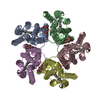 8r0oC  8r0pC M: map data used to model this data C: citing same article ( |
|---|---|
| Similar structure data |


-Links
 PDBj
PDBj


- Assembly
| Deposited unit | 
|
|---|---|
| 1 |
|
-Components
| #1: Protein | Mass: 35206.965 Da / Num. of mol.: 5 Source method: isolated from a genetically manipulated source Source: (gene. exp.) Cryobacterium levicorallinum (bacteria)Gene: E3O11_09160 / Production host: #2: Sugar | ChemComp-LMT /   Type: D-saccharide / Mass: 510.615 Da / Num. of mol.: 5 / Source method: obtained synthetically / Formula: C24H46O11 / Comment: detergent*YM Type: D-saccharide / Mass: 510.615 Da / Num. of mol.: 5 / Source method: obtained synthetically / Formula: C24H46O11 / Comment: detergent*YM#3: Chemical | ChemComp-RET /   Mass: 284.436 Da / Num. of mol.: 5 Mass: 284.436 Da / Num. of mol.: 5Source method: isolated from a genetically manipulated source Formula: C20H28O Source: (gene. exp.) Cryobacterium levicorallinum (bacteria)Gene: E3O11_09160 / Production host: #4: Chemical | ChemComp-LFA / |   Mass: 282.547 Da / Num. of mol.: 1 / Source method: isolated from a natural source / Formula: C20H42 Mass: 282.547 Da / Num. of mol.: 1 / Source method: isolated from a natural source / Formula: C20H42#5: Water | ChemComp-HOH / |  Mass: 18.015 Da / Num. of mol.: 78 / Source method: isolated from a natural source / Formula: H2O Mass: 18.015 Da / Num. of mol.: 78 / Source method: isolated from a natural source / Formula: H2OHas ligand of interest | Y | Has protein modification | Y | |
|---|
-Experimental details
-Experiment
| Experiment | Method: ELECTRON MICROSCOPY |
|---|---|
| EM experiment | Aggregation state: PARTICLE / 3D reconstruction method: single particle reconstruction |
- Sample preparation
| Component | Name: Pentameric form of the microbial rhodopsin CryoR1 / Type: COMPLEX Details: Solubilized in DDM, pH 10.5, ground state of CryoR1 Entity ID: #1 / Source: RECOMBINANT |
|---|---|
| Molecular weight | Value: 0.182 MDa / Experimental value: NO |
| Source (natural) | Organism: Cryobacterium levicorallinum (bacteria) |
| Source (recombinant) | Organism: |
| Buffer solution | pH: 10.5 |
| Specimen | Conc.: 7 mg/ml / Embedding applied: NO / Shadowing applied: NO / Staining applied: NO / Vitrification applied: YES |
| Specimen support | Grid material: GOLD / Grid mesh size: 300 divisions/in. / Grid type: Quantifoil R1.2/1.3 |
| Vitrification | Cryogen name: ETHANE |
- Electron microscopy imaging
| Experimental equipment |  Model: Titan Krios / Image courtesy: FEI Company |
|---|---|
| Microscopy | Model: TFS KRIOS |
| Electron gun | Electron source:  FIELD EMISSION GUN / Accelerating voltage: 300 kV / Illumination mode: FLOOD BEAM FIELD EMISSION GUN / Accelerating voltage: 300 kV / Illumination mode: FLOOD BEAM |
| Electron lens | Mode: OTHER / Nominal defocus max: 2200 nm / Nominal defocus min: 1200 nm / Cs: 2.7 mm |
| Image recording | Electron dose: 50 e/Å2 / Film or detector model: GATAN K3 (6k x 4k) |
- Processing
| EM software |
| ||||||||||||||||||||||||||||||||||||||||||||||||||||||||||||||||||||||||||||||||||||||||||||||||||||||||||
|---|---|---|---|---|---|---|---|---|---|---|---|---|---|---|---|---|---|---|---|---|---|---|---|---|---|---|---|---|---|---|---|---|---|---|---|---|---|---|---|---|---|---|---|---|---|---|---|---|---|---|---|---|---|---|---|---|---|---|---|---|---|---|---|---|---|---|---|---|---|---|---|---|---|---|---|---|---|---|---|---|---|---|---|---|---|---|---|---|---|---|---|---|---|---|---|---|---|---|---|---|---|---|---|---|---|---|---|
| CTF correction | Type: PHASE FLIPPING AND AMPLITUDE CORRECTION | ||||||||||||||||||||||||||||||||||||||||||||||||||||||||||||||||||||||||||||||||||||||||||||||||||||||||||
| Symmetry | Point symmetry: C5 (5 fold cyclic) | ||||||||||||||||||||||||||||||||||||||||||||||||||||||||||||||||||||||||||||||||||||||||||||||||||||||||||
| 3D reconstruction | Resolution: 2.7 Å / Resolution method: FSC 0.143 CUT-OFF / Num. of particles: 200653 / Symmetry type: POINT | ||||||||||||||||||||||||||||||||||||||||||||||||||||||||||||||||||||||||||||||||||||||||||||||||||||||||||
| Refinement | Resolution: 2.7→101.16 Å / Cor.coef. Fo:Fc: 0.598 / SU B: 10.55 / SU ML: 0.199 / ESU R: 0.537 Stereochemistry target values: MAXIMUM LIKELIHOOD WITH PHASES Details: HYDROGENS HAVE BEEN USED IF PRESENT IN THE INPUT
| ||||||||||||||||||||||||||||||||||||||||||||||||||||||||||||||||||||||||||||||||||||||||||||||||||||||||||
| Solvent computation | Solvent model: PARAMETERS FOR MASK CACLULATION | ||||||||||||||||||||||||||||||||||||||||||||||||||||||||||||||||||||||||||||||||||||||||||||||||||||||||||
| Displacement parameters | Biso mean: 46.352 Å2 | ||||||||||||||||||||||||||||||||||||||||||||||||||||||||||||||||||||||||||||||||||||||||||||||||||||||||||
| Refinement step | Cycle: 1 / Total: 10921 | ||||||||||||||||||||||||||||||||||||||||||||||||||||||||||||||||||||||||||||||||||||||||||||||||||||||||||
| Refine LS restraints |
|