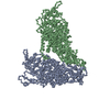
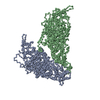
Journal: PLoS Pathog / Year: 2024 Title: High-resolution comparative atomic structures of two Giardiavirus prototypes infecting G. duodenalis parasite. Authors: Han Wang / Gianluca Marucci / Anna Munke / Mohammad Maruf Hassan / Marco Lalle / Kenta Okamoto / Abstract: The Giardia lamblia virus (GLV) is a non-enveloped icosahedral dsRNA and endosymbiont virus that infects the zoonotic protozoan parasite Giardia duodenalis (syn. G. lamblia, G. intestinalis), which ...The Giardia lamblia virus (GLV) is a non-enveloped icosahedral dsRNA and endosymbiont virus that infects the zoonotic protozoan parasite Giardia duodenalis (syn. G. lamblia, G. intestinalis), which is a pathogen of mammals, including humans. Elucidating the transmission mechanism of GLV is crucial for gaining an in-depth understanding of the virulence of the virus in G. duodenalis. GLV belongs to the family Totiviridae, which infects yeast and protozoa intracellularly; however, it also transmits extracellularly, similar to the phylogenetically, distantly related toti-like viruses that infect multicellular hosts. The GLV capsid structure is extensively involved in the longstanding discussion concerning extracellular transmission in Totiviridae and toti-like viruses. Hence, this study constructed the first high-resolution comparative atomic models of two GLV strains, namely GLV-HP and GLV-CAT, which showed different intracellular localization and virulence phenotypes, using cryogenic electron microscopy single-particle analysis. The atomic models of the GLV capsids presented swapped C-terminal extensions, extra surface loops, and a lack of cap-snatching pockets, similar to those of toti-like viruses. However, their open pores and absence of the extra crown protein resemble those of other yeast and protozoan Totiviridae viruses, demonstrating the essential structures for extracellular cell-to-cell transmission. The structural comparison between GLV-HP and GLV-CAT indicates the first evidence of critical structural motifs for the transmission and virulence of GLV in G. duodenalis.
History
Deposition
Oct 31, 2023
Deposition site: PDBE / Processing site: PDBE
Revision 1.0
Apr 3, 2024
Provider: repository / Type: Initial release
Revision 1.0
Apr 3, 2024
Data content type: EM metadata / Data content type: EM metadata / Provider: repository / Type: Initial release
Revision 1.0
Apr 3, 2024
Data content type: Half map / Part number: 1 / Data content type: Half map / Provider: repository / Type: Initial release
Revision 1.0
Apr 3, 2024
Data content type: Half map / Part number: 2 / Data content type: Half map / Provider: repository / Type: Initial release
Revision 1.0
Apr 3, 2024
Data content type: Image / Data content type: Image / Provider: repository / Type: Initial release
Revision 1.0
Apr 3, 2024
Data content type: Primary map / Data content type: Primary map / Provider: repository / Type: Initial release
Revision 1.0
Apr 3, 2024
Data content type: Half map / Part number: 1 / Data content type: Half map / Provider: repository / Type: Initial release
Revision 1.0
Apr 3, 2024
Data content type: Half map / Part number: 2 / Data content type: Half map / Provider: repository / Type: Initial release
Revision 1.0
Apr 3, 2024
Data content type: Image / Data content type: Image / Provider: repository / Type: Initial release
Revision 1.0
Apr 3, 2024
Data content type: Primary map / Data content type: Primary map / Provider: repository / Type: Initial release
Data content type: EM metadata / Data content type: EM metadata / EM metadata / Group: Data processing / Experimental summary / Data content type: EM metadata / EM metadata / Category: em_admin / em_software / Data content type: EM metadata / EM metadata / Item: _em_admin.last_update / _em_software.name
Mass: 103481.320 Da / Num. of mol.: 2 / Source method: isolated from a natural source / Source: (natural) Giardia lamblia virus / References: UniProt: A0A8F6AHR5
Has protein modification
N
-
Experimental details
-
Experiment
Experiment
Method: ELECTRON MICROSCOPY
EM experiment
Aggregation state: PARTICLE / 3D reconstruction method: single particle reconstruction
-
Sample preparation
Component
Name: Giardia lamblia virus / Type: VIRUS Details: GLV isolated from Giardia duodenalis isolates HP-1 from a human patient. Entity ID: all / Source: NATURAL
Molecular weight
Experimental value: NO
Source (natural)
Organism: Giardia lamblia virus / Strain: HP-1
Details of virus
Empty: NO / Enveloped: NO / Isolate: STRAIN / Type: VIRION
In the structure databanks used in Yorodumi, some data are registered as the other names, "COVID-19 virus" and "2019-nCoV". Here are the details of the virus and the list of structure data.
Jan 31, 2019. EMDB accession codes are about to change! (news from PDBe EMDB page)
EMDB accession codes are about to change! (news from PDBe EMDB page)
The allocation of 4 digits for EMDB accession codes will soon come to an end. Whilst these codes will remain in use, new EMDB accession codes will include an additional digit and will expand incrementally as the available range of codes is exhausted. The current 4-digit format prefixed with “EMD-” (i.e. EMD-XXXX) will advance to a 5-digit format (i.e. EMD-XXXXX), and so on. It is currently estimated that the 4-digit codes will be depleted around Spring 2019, at which point the 5-digit format will come into force.
The EM Navigator/Yorodumi systems omit the EMD- prefix.
Related info.:Q: What is EMD? / ID/Accession-code notation in Yorodumi/EM Navigator
Yorodumi is a browser for structure data from EMDB, PDB, SASBDB, etc.
This page is also the successor to EM Navigator detail page, and also detail information page/front-end page for Omokage search.
The word "yorodu" (or yorozu) is an old Japanese word meaning "ten thousand". "mi" (miru) is to see.
Related info.:EMDB / PDB / SASBDB / Comparison of 3 databanks / Yorodumi Search / Aug 31, 2016. New EM Navigator & Yorodumi / Yorodumi Papers / Jmol/JSmol / Function and homology information / Changes in new EM Navigator and Yorodumi
 Movie
Movie Controller


 Open data
 Basic information
 Components
 Keywords
 Function and homology information
 Giardia lamblia virus
 Authors
 Sweden,
  Norway, 3items
  Citation

 Structure visualization
 Downloads & links
 Other downloads

 PDBj
PDBj Assembly
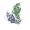
 Sample preparation
 Electron microscopy imaging
 FIELD EMISSION GUN / Accelerating voltage: 300 kV / Illumination mode: FLOOD BEAM
 Processing
