Journal: FEBS J / Year: 2024 Title: Structural characterization of the (deoxy)hypusination in Trichomonas vaginalis questions the bifunctionality of deoxyhypusine synthase. Authors: Elżbieta Wątor / Piotr Wilk / Paweł Kochanowski / Przemysław Grudnik / Abstract: Trichomonas vaginalis, the causative agent of trichomoniasis, is a prevalent anaerobic protozoan parasite responsible for the most common nonviral sexually transmitted infection globally. While ...Trichomonas vaginalis, the causative agent of trichomoniasis, is a prevalent anaerobic protozoan parasite responsible for the most common nonviral sexually transmitted infection globally. While metronidazole and its derivatives are approved drugs for this infection, rising resistance necessitates the exploration of new antiparasitic therapies. Protein posttranslational modifications (PTMs) play crucial roles in cellular processes, and among them, hypusination, involving eukaryotic translation factor 5A (eIF5A), has profound implications. Despite extensive studies in various organisms, the role of hypusination in T. vaginalis and its potential impact on parasite biology and pathogenicity remain poorly understood. This study aims to unravel the structural basis of the hypusination pathway in T. vaginalis using X-ray crystallography and cryo-electron microscopy. The results reveal high structural homology between T. vaginalis and human orthologs, providing insights into the molecular architecture of eIF5A and deoxyhypusine synthase (DHS) and their interaction. Contrary to previous suggestions of bifunctionality, our analyses indicate that the putative hydroxylation site in tvDHS is nonfunctional, and biochemical assays demonstrate exclusive deoxyhypusination capability. These findings challenge the notion of tvDHS functioning as both deoxyhypusine synthase and hydroxylase. The study enhances understanding of the hypusination pathway in T. vaginalis, shedding light on its functional relevance and potential as a drug target, and contributing to the development of novel therapeutic strategies against trichomoniasis.
In the structure databanks used in Yorodumi, some data are registered as the other names, "COVID-19 virus" and "2019-nCoV". Here are the details of the virus and the list of structure data.
Jan 31, 2019. EMDB accession codes are about to change! (news from PDBe EMDB page)
EMDB accession codes are about to change! (news from PDBe EMDB page)
The allocation of 4 digits for EMDB accession codes will soon come to an end. Whilst these codes will remain in use, new EMDB accession codes will include an additional digit and will expand incrementally as the available range of codes is exhausted. The current 4-digit format prefixed with “EMD-” (i.e. EMD-XXXX) will advance to a 5-digit format (i.e. EMD-XXXXX), and so on. It is currently estimated that the 4-digit codes will be depleted around Spring 2019, at which point the 5-digit format will come into force.
The EM Navigator/Yorodumi systems omit the EMD- prefix.
Related info.:Q: What is EMD? / ID/Accession-code notation in Yorodumi/EM Navigator
Yorodumi is a browser for structure data from EMDB, PDB, SASBDB, etc.
This page is also the successor to EM Navigator detail page, and also detail information page/front-end page for Omokage search.
The word "yorodu" (or yorozu) is an old Japanese word meaning "ten thousand". "mi" (miru) is to see.
Related info.:EMDB / PDB / SASBDB / Comparison of 3 databanks / Yorodumi Search / Aug 31, 2016. New EM Navigator & Yorodumi / Yorodumi Papers / Jmol/JSmol / Function and homology information / Changes in new EM Navigator and Yorodumi
 Movie
Movie Controller

 Yorodumi
 Open data
 Basic information
 Components
 Keywords
 Function and homology information
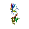
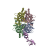
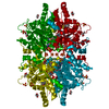
 Trichomonas vaginalis (eukaryote)
 X-RAY DIFFRACTION /
  Authors
 Poland, 2items
  Citation
 Structure visualization
 Downloads & links
 Other downloads
 PDBj
PDBj

 Assembly


 Mass: 663.425 Da / Num. of mol.: 1 / Source method: obtained synthetically / Formula: C21H27N7O14P2 / Feature type: SUBJECT OF INVESTIGATION / Comment: NAD*YM
 Mass: 96.063 Da / Num. of mol.: 7 / Source method: obtained synthetically / Formula: SO4
 Mass: 92.094 Da / Num. of mol.: 4 / Source method: obtained synthetically / Formula: C3H8O3
 Mass: 18.015 Da / Num. of mol.: 400 / Source method: isolated from a natural source / Formula: H2O
 Sample preparation
 / Beamline: 14.1 / Wavelength: 0.9184 Å
 Processing