 Movie
Movie Controller


+ Open data

- Basic information
| Entry | Database: PDB / ID: 8qxq | |||||||||
|---|---|---|---|---|---|---|---|---|---|---|
| Title | PsiM in complex with SAH and psilocybin | |||||||||
 Components Components | Psilocybin synthase | |||||||||
 Keywords Keywords | TRANSFERASE / Methyltransferase | |||||||||
| Function / homology |  Function and homology information Function and homology informationpsilocybin synthase / 4-hydroxytryptamine 4-phosphate methyltransferase activity / psilocybin biosynthetic process / rRNA base methylation / Transferases; Transferring one-carbon groups; Methyltransferases / nucleus Similarity search - Function | |||||||||
| Biological species |  Psilocybe cubensis (magic mushroom) Psilocybe cubensis (magic mushroom) | |||||||||
| Method |  X-RAY DIFFRACTION / SYNCHROTRON / FOURIER SYNTHESIS / Resolution: 0.94 Å X-RAY DIFFRACTION / SYNCHROTRON / FOURIER SYNTHESIS / Resolution: 0.94 Å | |||||||||
 Authors Authors | Werten, S. / Hudspeth, J. / Rupp, B. | |||||||||
| Funding support |  Austria, 1items Austria, 1items
| |||||||||
 Citation Citation | Journal: Nat Commun / Year: 2024 Title: Methyl transfer in psilocybin biosynthesis. Authors: Hudspeth, J. / Rogge, K. / Dorner, S. / Mull, M. / Hoffmeister, D. / Rupp, B. / Werten, S. | |||||||||
| History |
|
- Structure visualization
| Structure viewer | Molecule: MolmilJmol/JSmol |
|---|
- Downloads & links
-Download
| PDBx/mmCIF format | 8qxq.cif.gz | 240.8 KB | Display | PDBx/mmCIF format |
|---|---|---|---|---|
| PDB format | pdb8qxq.ent.gz | 186.6 KB | Display | PDB format |
| PDBx/mmJSON format | 8qxq.json.gz | Tree view | PDBx/mmJSON format | |
| Others |  Other downloads Other downloads |
-Validation report
| Summary document | 8qxq_validation.pdf.gz | 1.4 MB | Display | wwPDB validaton report |
|---|---|---|---|---|
| Full document | 8qxq_full_validation.pdf.gz | 1.4 MB | Display | |
| Data in XML | 8qxq_validation.xml.gz | 21.3 KB | Display | |
| Data in CIF | 8qxq_validation.cif.gz | 32.3 KB | Display | |
| Arichive directory | https://data.pdbj.org/pub/pdb/validation_reports/qx/8qxqftp://data.pdbj.org/pub/pdb/validation_reports/qx/8qxq | HTTPS FTP |
-Related structure data
| Related structure data | 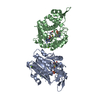 8pb3C  8pb4C 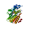 8pb5C 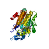 8pb6C  8pb7C 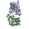 8pb8C C: citing same article ( |
|---|---|
| Similar structure data |
-Links
 PDBj
PDBj- Assembly
| Deposited unit | 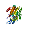
| ||||||||||||
|---|---|---|---|---|---|---|---|---|---|---|---|---|---|
| 1 |
| ||||||||||||
| Unit cell |
|
-Components
| #1: Protein | Mass: 35889.578 Da / Num. of mol.: 1 Source method: isolated from a genetically manipulated source Source: (gene. exp.) Psilocybe cubensis (magic mushroom) / Gene: psiM / Plasmid: pET-28a / Production host:  References: UniProt: P0DPA9, Transferases; Transferring one-carbon groups; Methyltransferases | ||||||
|---|---|---|---|---|---|---|---|
| #2: Chemical | ChemComp-SAH /   Mass: 384.411 Da / Num. of mol.: 1 / Source method: obtained synthetically / Formula: C14H20N6O5S / Feature type: SUBJECT OF INVESTIGATION Mass: 384.411 Da / Num. of mol.: 1 / Source method: obtained synthetically / Formula: C14H20N6O5S / Feature type: SUBJECT OF INVESTIGATION | ||||||
| #3: Chemical | ChemComp-X8Q / Mass: 284.248 Da / Num. of mol.: 1 / Source method: obtained synthetically / Formula: C12H17N2O4P / Feature type: SUBJECT OF INVESTIGATION | ||||||
| #4: Chemical |   Mass: 35.453 Da / Num. of mol.: 2 / Source method: obtained synthetically / Formula: Cl Mass: 35.453 Da / Num. of mol.: 2 / Source method: obtained synthetically / Formula: Cl#5: Water | ChemComp-HOH / |  Mass: 18.015 Da / Num. of mol.: 451 / Source method: isolated from a natural source / Formula: H2O Mass: 18.015 Da / Num. of mol.: 451 / Source method: isolated from a natural source / Formula: H2OHas ligand of interest | Y | Has protein modification | N | |
-Experimental details
-Experiment
| Experiment | Method: X-RAY DIFFRACTION / Number of used crystals: 1 |
|---|
- Sample preparation
| Crystal | Density Matthews: 2.27 Å3/Da / Density % sol: 45.78 % |
|---|---|
| Crystal grow | Temperature: 277 K / Method: vapor diffusion, hanging drop / pH: 8.5 / Details: 100 mM Tris/HCl pH 8.5, 20% PEG 8000, 200 mM MgCl2 |
-Data collection
| Diffraction | Mean temperature: 100 K / Serial crystal experiment: N |
|---|---|
| Diffraction source | Source: SYNCHROTRON / Site: ESRF  / Beamline: ID23-1 / Wavelength: 0.7293 Å / Beamline: ID23-1 / Wavelength: 0.7293 Å |
| Detector | Type: DECTRIS EIGER2 X CdTe 16M / Detector: PIXEL / Date: Jun 29, 2023 / Details: Toroidal mirror |
| Radiation | Monochromator: Si(111) / Protocol: SINGLE WAVELENGTH / Monochromatic (M) / Laue (L): M / Scattering type: x-ray |
| Radiation wavelength | Wavelength: 0.7293 Å / Relative weight: 1 |
| Reflection | Resolution: 0.94→15.44 Å / Num. obs: 211345 / % possible obs: 99.81 % / Redundancy: 26.8 % / Biso Wilson estimate: 11.17 Å2 / CC1/2: 0.998 / CC star: 1 / Rmerge(I) obs: 0.1082 / Rpim(I) all: 0.0212 / Rrim(I) all: 0.1103 / Net I/σ(I): 13.36 |
| Reflection shell | Resolution: 0.94→0.9736 Å / Redundancy: 26.9 % / Rmerge(I) obs: 4.584 / Mean I/σ(I) obs: 0.53 / Num. unique obs: 20765 / CC1/2: 0.383 / CC star: 0.745 / Rpim(I) all: 0.8985 / Rrim(I) all: 4.673 / % possible all: 99.16 |
- Processing
| Software |
| ||||||||||||||||||||||||||||||||||||||||||||||||||||||||||||||||||||||||||||||||||||||||||||||||||||||||||||||||||||||||||||||||||||||||||||||||||||||||||||||||||||||||
|---|---|---|---|---|---|---|---|---|---|---|---|---|---|---|---|---|---|---|---|---|---|---|---|---|---|---|---|---|---|---|---|---|---|---|---|---|---|---|---|---|---|---|---|---|---|---|---|---|---|---|---|---|---|---|---|---|---|---|---|---|---|---|---|---|---|---|---|---|---|---|---|---|---|---|---|---|---|---|---|---|---|---|---|---|---|---|---|---|---|---|---|---|---|---|---|---|---|---|---|---|---|---|---|---|---|---|---|---|---|---|---|---|---|---|---|---|---|---|---|---|---|---|---|---|---|---|---|---|---|---|---|---|---|---|---|---|---|---|---|---|---|---|---|---|---|---|---|---|---|---|---|---|---|---|---|---|---|---|---|---|---|---|---|---|---|---|---|---|---|
| Refinement | Method to determine structure: FOURIER SYNTHESIS / Resolution: 0.94→15.44 Å / SU ML: 0.1239 / Cross valid method: FREE R-VALUE / σ(F): 1.33 / Phase error: 16.1338 Stereochemistry target values: GeoStd + Monomer Library + CDL v1.2
| ||||||||||||||||||||||||||||||||||||||||||||||||||||||||||||||||||||||||||||||||||||||||||||||||||||||||||||||||||||||||||||||||||||||||||||||||||||||||||||||||||||||||
| Solvent computation | Shrinkage radii: 0.9 Å / VDW probe radii: 1.1 Å / Solvent model: FLAT BULK SOLVENT MODEL | ||||||||||||||||||||||||||||||||||||||||||||||||||||||||||||||||||||||||||||||||||||||||||||||||||||||||||||||||||||||||||||||||||||||||||||||||||||||||||||||||||||||||
| Displacement parameters | Biso mean: 16.66 Å2 | ||||||||||||||||||||||||||||||||||||||||||||||||||||||||||||||||||||||||||||||||||||||||||||||||||||||||||||||||||||||||||||||||||||||||||||||||||||||||||||||||||||||||
| Refinement step | Cycle: LAST / Resolution: 0.94→15.44 Å
| ||||||||||||||||||||||||||||||||||||||||||||||||||||||||||||||||||||||||||||||||||||||||||||||||||||||||||||||||||||||||||||||||||||||||||||||||||||||||||||||||||||||||
| Refine LS restraints |
| ||||||||||||||||||||||||||||||||||||||||||||||||||||||||||||||||||||||||||||||||||||||||||||||||||||||||||||||||||||||||||||||||||||||||||||||||||||||||||||||||||||||||
| LS refinement shell |
|
