 Movie
Movie Controller

[English] 日本語
 Yorodumi
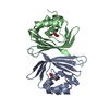
- PDB-8qx5: Helical Carotenoid Protein 4 (HCP4) from Anabaena with bound Cant... -
+ Open data

- Basic information
| Entry | Database: PDB / ID: 8qx5 | ||||||
|---|---|---|---|---|---|---|---|
| Title | Helical Carotenoid Protein 4 (HCP4) from Anabaena with bound Canthaxanthin | ||||||
 Components Components | Orange carotenoid-binding domain-containing protein | ||||||
 Keywords Keywords | PHOTOSYNTHESIS / Photoprotection / soluble proteins / Orange carotenoid protein / Cyanobacteria | ||||||
| Function / homology |  Function and homology information Function and homology informationlight absorption / phycobilisome / chloride ion binding / plasma membrane-derived thylakoid membrane Similarity search - Function | ||||||
| Biological species |  Cyanobacteriota (cyanobacteria) Cyanobacteriota (cyanobacteria) | ||||||
| Method |  X-RAY DIFFRACTION / SYNCHROTRON / MOLECULAR REPLACEMENT / Resolution: 1.9 Å X-RAY DIFFRACTION / SYNCHROTRON / MOLECULAR REPLACEMENT / Resolution: 1.9 Å | ||||||
 Authors Authors | Sklyar, J. / Wilson, A. / Kirilovsky, D. / Adir, N. | ||||||
| Funding support |  Israel, 1items Israel, 1items
| ||||||
 Citation Citation | Journal: Int.J.Biol.Macromol. / Year: 2024 Title: Insights into energy quenching mechanisms and carotenoid uptake by orange carotenoid protein homologs: HCP4 and CTDH. Authors: Sklyar, J. / Wilson, A. / Kirilovsky, D. / Adir, N. | ||||||
| History |
|
- Structure visualization
| Structure viewer | Molecule: MolmilJmol/JSmol |
|---|
- Downloads & links
-Download
| PDBx/mmCIF format | 8qx5.cif.gz | 97.8 KB | Display | PDBx/mmCIF format |
|---|---|---|---|---|
| PDB format | pdb8qx5.ent.gz | 61 KB | Display | PDB format |
| PDBx/mmJSON format | 8qx5.json.gz | Tree view | PDBx/mmJSON format | |
| Others |  Other downloads Other downloads |
-Validation report
| Arichive directory | https://data.pdbj.org/pub/pdb/validation_reports/qx/8qx5ftp://data.pdbj.org/pub/pdb/validation_reports/qx/8qx5 | HTTPS FTP |
|---|
-Related structure data
-Links
 PDBj
PDBj- Assembly
| Deposited unit | 
| ||||||||||||
|---|---|---|---|---|---|---|---|---|---|---|---|---|---|
| 1 | 
| ||||||||||||
| 2 | 
| ||||||||||||
| Unit cell |
| ||||||||||||
| Components on special symmetry positions |
|
-Components
| #1: Protein | Mass: 19444.033 Da / Num. of mol.: 2 Source method: isolated from a genetically manipulated source Source: (gene. exp.) Cyanobacteriota (cyanobacteria) / Strain: Anabaena / Gene: all4941 / Production host: #2: Chemical |   Mass: 564.840 Da / Num. of mol.: 2 / Source method: obtained synthetically / Formula: C40H52O2 / Feature type: SUBJECT OF INVESTIGATION Mass: 564.840 Da / Num. of mol.: 2 / Source method: obtained synthetically / Formula: C40H52O2 / Feature type: SUBJECT OF INVESTIGATION#3: Water | ChemComp-HOH / |  Mass: 18.015 Da / Num. of mol.: 186 / Source method: isolated from a natural source / Formula: H2O Mass: 18.015 Da / Num. of mol.: 186 / Source method: isolated from a natural source / Formula: H2OHas ligand of interest | Y | |
|---|
-Experimental details
-Experiment
| Experiment | Method: X-RAY DIFFRACTION / Number of used crystals: 1 |
|---|
- Sample preparation
| Crystal | Density Matthews: 2.09 Å3/Da / Density % sol: 41.12 % |
|---|---|
| Crystal grow | Temperature: 293 K / Method: vapor diffusion, hanging drop / pH: 5.6 Details: 0.095M sodium citrate tribasic dihydrate pH=5.6, 19% v/v 2-propanol, 19% w/v PEG 4000, 5% glycerol |
-Data collection
| Diffraction | Mean temperature: 100 K / Serial crystal experiment: N |
|---|---|
| Diffraction source | Source: SYNCHROTRON / Site: Diamond  / Beamline: I04 / Wavelength: 0.9537 Å / Beamline: I04 / Wavelength: 0.9537 Å |
| Detector | Type: DECTRIS EIGER2 XE 16M / Detector: PIXEL / Date: Jul 27, 2023 |
| Radiation | Protocol: SINGLE WAVELENGTH / Monochromatic (M) / Laue (L): M / Scattering type: x-ray |
| Radiation wavelength | Wavelength: 0.9537 Å / Relative weight: 1 |
| Reflection | Resolution: 1.9→29.73 Å / Num. obs: 27148 / % possible obs: 99.9 % / Redundancy: 26.2 % / CC1/2: 1 / Rmerge(I) obs: 0.089 / Rpim(I) all: 0.024 / Rrim(I) all: 0.092 / Net I/σ(I): 25 |
| Reflection shell | Resolution: 1.9→1.94 Å / Num. unique obs: 1679 / CC1/2: 0.955 |
- Processing
| Software |
| |||||||||||||||||||||||||||||||||||||||||||||||||||||||||||||||||||||||||||||
|---|---|---|---|---|---|---|---|---|---|---|---|---|---|---|---|---|---|---|---|---|---|---|---|---|---|---|---|---|---|---|---|---|---|---|---|---|---|---|---|---|---|---|---|---|---|---|---|---|---|---|---|---|---|---|---|---|---|---|---|---|---|---|---|---|---|---|---|---|---|---|---|---|---|---|---|---|---|---|
| Refinement | Method to determine structure: MOLECULAR REPLACEMENT / Resolution: 1.9→29.73 Å / SU ML: 0.2205 / Cross valid method: FREE R-VALUE / Phase error: 22.1239 Stereochemistry target values: GeoStd + Monomer Library + CDL v1.2
| |||||||||||||||||||||||||||||||||||||||||||||||||||||||||||||||||||||||||||||
| Solvent computation | Shrinkage radii: 0.9 Å / VDW probe radii: 1.1 Å / Solvent model: FLAT BULK SOLVENT MODEL | |||||||||||||||||||||||||||||||||||||||||||||||||||||||||||||||||||||||||||||
| Refinement step | Cycle: LAST / Resolution: 1.9→29.73 Å
| |||||||||||||||||||||||||||||||||||||||||||||||||||||||||||||||||||||||||||||
| Refine LS restraints |
| |||||||||||||||||||||||||||||||||||||||||||||||||||||||||||||||||||||||||||||
| LS refinement shell |
|
