 Movie
Movie Controller

[English] 日本語
 Yorodumi
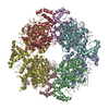
- PDB-8qwb: Crystal structure of citrate synthase from Methylophaga sulfidovorans -
+ Open data

- Basic information
| Entry | Database: PDB / ID: 8qwb | ||||||
|---|---|---|---|---|---|---|---|
| Title | Crystal structure of citrate synthase from Methylophaga sulfidovorans | ||||||
 Components Components | Citrate synthase | ||||||
 Keywords Keywords | TRANSFERASE / Citrate Synthase | ||||||
| Function / homology |  Function and homology information Function and homology information | ||||||
| Biological species |  Methylophaga sulfidovorans (bacteria) Methylophaga sulfidovorans (bacteria) | ||||||
| Method |  X-RAY DIFFRACTION / SYNCHROTRON / MOLECULAR REPLACEMENT / Resolution: 3.201 Å X-RAY DIFFRACTION / SYNCHROTRON / MOLECULAR REPLACEMENT / Resolution: 3.201 Å | ||||||
 Authors Authors | Mais, C.-N. / Bange, G. / Sendker, F.L. / Hochberg, G. | ||||||
| Funding support | 1items
| ||||||
 Citation Citation | Journal: bioRxiv / Year: 2024 Title: Frequent transitions in self-assembly across the evolution of a central metabolic enzyme. Authors: Franziska L Sendker / Tabea Schlotthauer / Christopher-Nils Mais / Yat Kei Lo / Mathias Girbig / Stefan Bohn / Thomas Heimerl / Daniel Schindler / Arielle Weinstein / Brain P Metzger / ...Authors: Franziska L Sendker / Tabea Schlotthauer / Christopher-Nils Mais / Yat Kei Lo / Mathias Girbig / Stefan Bohn / Thomas Heimerl / Daniel Schindler / Arielle Weinstein / Brain P Metzger / Joseph W Thornton / Arvind Pillai / Gert Bange / Jan M Schuller / Georg K A Hochberg /   Abstract: Many enzymes assemble into homomeric protein complexes comprising multiple copies of one protein. Because structural form is usually assumed to follow function in biochemistry, these assemblies are ...Many enzymes assemble into homomeric protein complexes comprising multiple copies of one protein. Because structural form is usually assumed to follow function in biochemistry, these assemblies are thought to evolve because they provide some functional advantage. In many cases, however, no specific advantage is known and, in some cases, quaternary structure varies among orthologs. This has led to the proposition that self-assembly may instead vary neutrally within protein families. The extent of such variation has been difficult to ascertain because quaternary structure has until recently been difficult to measure on large scales. Here, we employ mass photometry, phylogenetics, and structural biology to interrogate the evolution of homo-oligomeric assembly across the entire phylogeny of prokaryotic citrate synthases - an enzyme with a highly conserved function. We discover a menagerie of different assembly types that come and go over the course of evolution, including cases of parallel evolution and reversions from complex to simple assemblies. Functional experiments in vitro and in vivo indicate that evolutionary transitions between different assemblies do not strongly influence enzyme catalysis. Our work suggests that enzymes can wander relatively freely through a large space of possible assemblies and demonstrates the power of characterizing structure-function relationships across entire phylogenies. | ||||||
| History |
|
- Structure visualization
| Structure viewer | Molecule: MolmilJmol/JSmol |
|---|
- Downloads & links
-Download
| PDBx/mmCIF format | 8qwb.cif.gz | 1.4 MB | Display | PDBx/mmCIF format |
|---|---|---|---|---|
| PDB format | pdb8qwb.ent.gz | 1.2 MB | Display | PDB format |
| PDBx/mmJSON format | 8qwb.json.gz | Tree view | PDBx/mmJSON format | |
| Others |  Other downloads Other downloads |
-Validation report
| Arichive directory | https://data.pdbj.org/pub/pdb/validation_reports/qw/8qwbftp://data.pdbj.org/pub/pdb/validation_reports/qw/8qwb | HTTPS FTP |
|---|
-Related structure data

-Links
 PDBj
PDBj

- Assembly
| Deposited unit | 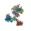
| ||||||||
|---|---|---|---|---|---|---|---|---|---|
| 1 | 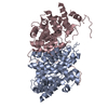
| ||||||||
| 2 | 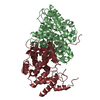
| ||||||||
| 3 | 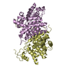
| ||||||||
| 4 | 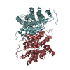
| ||||||||
| 5 | 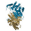
| ||||||||
| 6 | 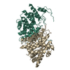
| ||||||||
| Unit cell |
|
-Components
| #1: Protein | Mass: 44767.852 Da / Num. of mol.: 12 Source method: isolated from a genetically manipulated source Source: (gene. exp.) Methylophaga sulfidovorans (bacteria) / Gene: SAMN04488079_11738 / Production host: |
|---|
-Experimental details
-Experiment
| Experiment | Method: X-RAY DIFFRACTION / Number of used crystals: 1 |
|---|
- Sample preparation
| Crystal | Density Matthews: 3.34 Å3/Da / Density % sol: 63.16 % |
|---|---|
| Crystal grow | Temperature: 293 K / Method: vapor diffusion, sitting drop Details: 1.0 M Lithium chloride, 0.1 M Citric Acid pH 5.0, 10% PEG 6000, 5mM oxaloacetic acid |
-Data collection
| Diffraction | Mean temperature: 100 K / Serial crystal experiment: N | ||||||||||||||||||||||||||||||||||||||||||||||||||||||||||||||||||||||||||||||||||||||||||||||||||||||||||||||||||||||||||||||
|---|---|---|---|---|---|---|---|---|---|---|---|---|---|---|---|---|---|---|---|---|---|---|---|---|---|---|---|---|---|---|---|---|---|---|---|---|---|---|---|---|---|---|---|---|---|---|---|---|---|---|---|---|---|---|---|---|---|---|---|---|---|---|---|---|---|---|---|---|---|---|---|---|---|---|---|---|---|---|---|---|---|---|---|---|---|---|---|---|---|---|---|---|---|---|---|---|---|---|---|---|---|---|---|---|---|---|---|---|---|---|---|---|---|---|---|---|---|---|---|---|---|---|---|---|---|---|---|
| Diffraction source | Source: SYNCHROTRON / Site: PETRA III, EMBL c/o DESY / Beamline: P13 (MX1) / Wavelength: 0.97625 Å | ||||||||||||||||||||||||||||||||||||||||||||||||||||||||||||||||||||||||||||||||||||||||||||||||||||||||||||||||||||||||||||||
| Detector | Type: DECTRIS EIGER X 16M / Detector: PIXEL / Date: Aug 29, 2020 | ||||||||||||||||||||||||||||||||||||||||||||||||||||||||||||||||||||||||||||||||||||||||||||||||||||||||||||||||||||||||||||||
| Radiation | Protocol: SINGLE WAVELENGTH / Monochromatic (M) / Laue (L): M / Scattering type: x-ray | ||||||||||||||||||||||||||||||||||||||||||||||||||||||||||||||||||||||||||||||||||||||||||||||||||||||||||||||||||||||||||||||
| Radiation wavelength | Wavelength: 0.97625 Å / Relative weight: 1 | ||||||||||||||||||||||||||||||||||||||||||||||||||||||||||||||||||||||||||||||||||||||||||||||||||||||||||||||||||||||||||||||
| Reflection | Resolution: 3.2→48.31 Å / Num. obs: 636593 / % possible obs: 98.9 % / Redundancy: 6.9 % / CC1/2: 0.998 / Rmerge(I) obs: 0.131 / Rrim(I) all: 0.142 / Net I/σ(I): 10.29 | ||||||||||||||||||||||||||||||||||||||||||||||||||||||||||||||||||||||||||||||||||||||||||||||||||||||||||||||||||||||||||||||
| Reflection shell |
|
- Processing
| Software |
| ||||||||||||||||
|---|---|---|---|---|---|---|---|---|---|---|---|---|---|---|---|---|---|
| Refinement | Method to determine structure: MOLECULAR REPLACEMENT / Resolution: 3.201→48.31 Å / Cross valid method: FREE R-VALUE
| ||||||||||||||||
| Refinement step | Cycle: LAST / Resolution: 3.201→48.31 Å
|
