 Movie
Movie Controller

[English] 日本語
 Yorodumi
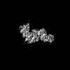
- PDB-8qvw: Cryo-EM structure of the peptide binding domain of human SRP68/72 -
+ Open data

- Basic information
| Entry | Database: PDB / ID: 8qvw | ||||||
|---|---|---|---|---|---|---|---|
| Title | Cryo-EM structure of the peptide binding domain of human SRP68/72 | ||||||
 Components Components |
| ||||||
 Keywords Keywords | TRANSLATION / Signal recognition particle / TPR / protein translocation | ||||||
| Function / homology |  Function and homology information Function and homology informationendoplasmic reticulum signal sequence receptor activity / signal recognition particle, endoplasmic reticulum targeting / signal recognition particle / signal recognition particle binding / 7S RNA binding / SRP-dependent cotranslational protein targeting to membrane / TPR domain binding / SRP-dependent cotranslational protein targeting to membrane / response to xenobiotic stimulus / ribosome ...endoplasmic reticulum signal sequence receptor activity / signal recognition particle, endoplasmic reticulum targeting / signal recognition particle / signal recognition particle binding / 7S RNA binding / SRP-dependent cotranslational protein targeting to membrane / TPR domain binding / SRP-dependent cotranslational protein targeting to membrane / response to xenobiotic stimulus / ribosome / protein domain specific binding / focal adhesion / nucleolus / endoplasmic reticulum / RNA binding / cytosol Similarity search - Function | ||||||
| Biological species |  Homo sapiens (human) Homo sapiens (human) | ||||||
| Method | ELECTRON MICROSCOPY / single particle reconstruction / cryo EM / Resolution: 3 Å | ||||||
 Authors Authors | Zhong, Y. / Feng, J. / Koh, A.F. / Kotecha, A. / Greber, B.J. / Ataide, S.F. | ||||||
| Funding support |  United Kingdom, 1items United Kingdom, 1items
| ||||||
 Citation Citation | Journal: Nucleic Acids Res / Year: 2024 Title: Cryo-EM structure of SRP68/72 reveals an extended dimerization domain with RNA-binding activity. Authors: Yichen Zhong / Junjie Feng / Adrian F Koh / Abhay Kotecha / Basil J Greber / Sandro F Ataide /   Abstract: The signal recognition particle (SRP) is a critical component in protein sorting pathways in all domains of life. Human SRP contains six proteins bound to the 7S RNA and their structures and ...The signal recognition particle (SRP) is a critical component in protein sorting pathways in all domains of life. Human SRP contains six proteins bound to the 7S RNA and their structures and functions have been mostly elucidated. The SRP68/72 dimer is the largest SRP component and is essential for SRP function. Although the structures of the SRP68/72 RNA binding and dimerization domains have been previously reported, the structure and function of large portions of the SRP68/72 dimer remain unknown. Here, we analyse full-length SRP68/72 using cryo-EM and report that SRP68/72 depend on each other for stability and form an extended dimerization domain. This newly observed dimerization domain is both a protein- and RNA-binding domain. Comparative analysis with current structural models suggests that this dimerization domain undergoes dramatic translocation upon SRP docking onto SRP receptor and eventually comes close to the Alu domain. We propose that the SRP68/72 dimerization domain functions by binding and detaching the Alu domain and SRP9/14 from the ribosomal surface, thus releasing elongation arrest upon docking onto the ER membrane. | ||||||
| History |
|
- Structure visualization
| Structure viewer | Molecule: MolmilJmol/JSmol |
|---|
- Downloads & links
-Download
| PDBx/mmCIF format | 8qvw.cif.gz | 125.3 KB | Display | PDBx/mmCIF format |
|---|---|---|---|---|
| PDB format | pdb8qvw.ent.gz | 85.1 KB | Display | PDB format |
| PDBx/mmJSON format | 8qvw.json.gz | Tree view | PDBx/mmJSON format | |
| Others |  Other downloads Other downloads |
-Validation report
| Arichive directory | https://data.pdbj.org/pub/pdb/validation_reports/qv/8qvwftp://data.pdbj.org/pub/pdb/validation_reports/qv/8qvw | HTTPS FTP |
|---|
-Related structure data
| Related structure data |  18674MC  8qvxC M: map data used to model this data C: citing same article ( |
|---|---|
| Similar structure data |
-Links
 PDBj
PDBj


- Assembly
| Deposited unit | 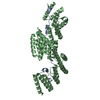
|
|---|---|
| 1 |
|
-Components
| #1: Protein | Mass: 66617.305 Da / Num. of mol.: 1 Source method: isolated from a genetically manipulated source Source: (gene. exp.) Homo sapiens (human) / Gene: SRP68 / Production host:  |
|---|---|
| #2: Protein | Mass: 74992.531 Da / Num. of mol.: 1 Source method: isolated from a genetically manipulated source Source: (gene. exp.) Homo sapiens (human) / Gene: SRP72 / Production host: |
-Experimental details
-Experiment
| Experiment | Method: ELECTRON MICROSCOPY |
|---|---|
| EM experiment | Aggregation state: PARTICLE / 3D reconstruction method: single particle reconstruction |
- Sample preparation
| Component | Name: SRP68/72 / Type: COMPLEX / Entity ID: all / Source: RECOMBINANT | ||||||||||||||||||||||||||||||
|---|---|---|---|---|---|---|---|---|---|---|---|---|---|---|---|---|---|---|---|---|---|---|---|---|---|---|---|---|---|---|---|
| Molecular weight | Value: 0.14 MDa / Experimental value: NO | ||||||||||||||||||||||||||||||
| Source (natural) | Organism: Homo sapiens (human) | ||||||||||||||||||||||||||||||
| Source (recombinant) | Organism: | ||||||||||||||||||||||||||||||
| Buffer solution | pH: 7.5 | ||||||||||||||||||||||||||||||
| Buffer component |
| ||||||||||||||||||||||||||||||
| Specimen | Conc.: 0.4 mg/ml / Embedding applied: NO / Shadowing applied: NO / Staining applied: NO / Vitrification applied: YES | ||||||||||||||||||||||||||||||
| Specimen support | Grid material: GOLD / Grid mesh size: 300 divisions/in. / Grid type: UltrAuFoil R1.2/1.3 | ||||||||||||||||||||||||||||||
| Vitrification | Instrument: FEI VITROBOT MARK IV / Cryogen name: ETHANE / Humidity: 100 % / Chamber temperature: 278 K |
- Electron microscopy imaging
| Experimental equipment |  Model: Titan Krios / Image courtesy: FEI Company |
|---|---|
| Microscopy | Model: TFS KRIOS |
| Electron gun | Electron source:  FIELD EMISSION GUN / Accelerating voltage: 300 kV / Illumination mode: FLOOD BEAM FIELD EMISSION GUN / Accelerating voltage: 300 kV / Illumination mode: FLOOD BEAM |
| Electron lens | Mode: BRIGHT FIELD / Calibrated magnification: 245614 X / Nominal defocus max: 2000 nm / Nominal defocus min: 500 nm / Cs: 2.7 mm / C2 aperture diameter: 50 µm / Alignment procedure: COMA FREE |
| Specimen holder | Cryogen: NITROGEN / Specimen holder model: FEI TITAN KRIOS AUTOGRID HOLDER |
| Image recording | Electron dose: 70 e/Å2 / Film or detector model: TFS FALCON 4i (4k x 4k) / Num. of grids imaged: 1 / Num. of real images: 18601 |
| EM imaging optics | Energyfilter name: TFS Selectris X / Energyfilter slit width: 10 eV |
- Processing
| EM software |
| |||||||||||||||||||||||||||||||||||||||||||||||||||||||
|---|---|---|---|---|---|---|---|---|---|---|---|---|---|---|---|---|---|---|---|---|---|---|---|---|---|---|---|---|---|---|---|---|---|---|---|---|---|---|---|---|---|---|---|---|---|---|---|---|---|---|---|---|---|---|---|---|
| Image processing | Details: Collected movies in EER format. | |||||||||||||||||||||||||||||||||||||||||||||||||||||||
| CTF correction | Type: PHASE FLIPPING AND AMPLITUDE CORRECTION | |||||||||||||||||||||||||||||||||||||||||||||||||||||||
| Symmetry | Point symmetry: C1 (asymmetric) | |||||||||||||||||||||||||||||||||||||||||||||||||||||||
| 3D reconstruction | Resolution: 3 Å / Resolution method: FSC 0.143 CUT-OFF / Num. of particles: 38514 / Algorithm: FOURIER SPACE / Details: BLUSH regularisation in RELION 5 used. / Num. of class averages: 1 / Symmetry type: POINT | |||||||||||||||||||||||||||||||||||||||||||||||||||||||
| Atomic model building | Protocol: RIGID BODY FIT / Space: REAL | |||||||||||||||||||||||||||||||||||||||||||||||||||||||
| Atomic model building | Source name: AlphaFold / Type: in silico model | |||||||||||||||||||||||||||||||||||||||||||||||||||||||
| Refine LS restraints |
|
