 Movie
Movie Controller

[English] 日本語
 Yorodumi
- PDB-8qt8: Crystal structure of human Sirt2 in complex with a TNFa-Myr analo... -
+ Open data

- Basic information
| Entry | Database: PDB / ID: 8qt8 | |||||||||
|---|---|---|---|---|---|---|---|---|---|---|
| Title | Crystal structure of human Sirt2 in complex with a TNFa-Myr analogue TNFn-34 | |||||||||
 Components Components |
| |||||||||
 Keywords Keywords | HYDROLASE / Super-slow substrate / Sirtuin 2 | |||||||||
| Function / homology |  Function and homology information Function and homology informationcellular response to caloric restriction / histone methacryllysine demethacrylase activity / histone benzoyllysine debenzoylase activity / negative regulation of oligodendrocyte progenitor proliferation / negative regulation of striated muscle tissue development / negative regulation of satellite cell differentiation / histone H4K16 deacetylase activity, NAD-dependent / positive regulation of attachment of spindle microtubules to kinetochore / NAD-dependent protein demyristoylase activity / NAD-dependent protein depalmitoylase activity ...cellular response to caloric restriction / histone methacryllysine demethacrylase activity / histone benzoyllysine debenzoylase activity / negative regulation of oligodendrocyte progenitor proliferation / negative regulation of striated muscle tissue development / negative regulation of satellite cell differentiation / histone H4K16 deacetylase activity, NAD-dependent / positive regulation of attachment of spindle microtubules to kinetochore / NAD-dependent protein demyristoylase activity / NAD-dependent protein depalmitoylase activity / peptidyl-lysine deacetylation / positive regulation of meiotic nuclear division / paranodal junction / lateral loop / mitotic nuclear membrane reassembly / regulation of exit from mitosis / tubulin deacetylase activity / regulation of phosphorylation / paranode region of axon / myelination in peripheral nervous system / tubulin deacetylation / negative regulation of peptidyl-threonine phosphorylation / Schmidt-Lanterman incisure / positive regulation of fatty acid biosynthetic process / regulation of myelination / protein acetyllysine N-acetyltransferase / NAD-dependent protein lysine deacetylase activity / histone deacetylase activity, NAD-dependent / rDNA heterochromatin formation / positive regulation of oocyte maturation / juxtaparanode region of axon / Initiation of Nuclear Envelope (NE) Reformation / protein deacetylation / chromatin silencing complex / meiotic spindle / histone deacetylase activity / protein lysine deacetylase activity / response to redox state / negative regulation of fat cell differentiation / positive regulation of DNA binding / histone acetyltransferase binding / negative regulation of reactive oxygen species metabolic process / positive regulation of cell division / NAD+ poly-ADP-ribosyltransferase activity / NAD+ binding / subtelomeric heterochromatin formation / positive regulation of execution phase of apoptosis / glial cell projection / lipid catabolic process / heterochromatin / centriole / cellular response to epinephrine stimulus / Transferases; Acyltransferases; Transferring groups other than aminoacyl groups / substantia nigra development / negative regulation of autophagy / epigenetic regulation of gene expression / ubiquitin binding / negative regulation of protein catabolic process / meiotic cell cycle / mitotic spindle / spindle / histone deacetylase binding / autophagy / myelin sheath / positive regulation of proteasomal ubiquitin-dependent protein catabolic process / chromosome / growth cone / cellular response to oxidative stress / heterochromatin formation / midbody / cellular response to hypoxia / DNA-binding transcription factor binding / microtubule / proteasome-mediated ubiquitin-dependent protein catabolic process / perikaryon / chromosome, telomeric region / regulation of cell cycle / innate immune response / cell division / negative regulation of DNA-templated transcription / centrosome / chromatin binding / nucleolus / perinuclear region of cytoplasm / negative regulation of transcription by RNA polymerase II / positive regulation of transcription by RNA polymerase II / mitochondrion / DNA-templated transcription / nucleoplasm / zinc ion binding / nucleus / cytosol / cytoplasm Similarity search - Function | |||||||||
| Biological species |  Homo sapiens (human) Homo sapiens (human) | |||||||||
| Method |  X-RAY DIFFRACTION / SYNCHROTRON / MOLECULAR REPLACEMENT / Resolution: 1.65 Å X-RAY DIFFRACTION / SYNCHROTRON / MOLECULAR REPLACEMENT / Resolution: 1.65 Å | |||||||||
 Authors Authors | Friedrich, F. / Kalbas, D. / Meleshin, M. / Einsle, O. / Schutkowski, M. / Jung, M. | |||||||||
| Funding support |  Germany, 2items Germany, 2items
| |||||||||
 Citation Citation | Journal: To Be Published Title: New Super-Slow Substrates as novel Sirtuin-Inhibitors Authors: Schutkowski, M. / Kalbas, D. / Einsle, O. / Schutkowski, M. / Jung, M. | |||||||||
| History |
|
- Structure visualization
| Structure viewer | Molecule: MolmilJmol/JSmol |
|---|
- Downloads & links
-Download
| PDBx/mmCIF format | 8qt8.cif.gz | 143.2 KB | Display | PDBx/mmCIF format |
|---|---|---|---|---|
| PDB format | pdb8qt8.ent.gz | 108.9 KB | Display | PDB format |
| PDBx/mmJSON format | 8qt8.json.gz | Tree view | PDBx/mmJSON format | |
| Others |  Other downloads Other downloads |
-Validation report
| Arichive directory | https://data.pdbj.org/pub/pdb/validation_reports/qt/8qt8ftp://data.pdbj.org/pub/pdb/validation_reports/qt/8qt8 | HTTPS FTP |
|---|
-Related structure data
| Related structure data |  8qt0C 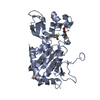 8qt1C  8qt2C  8qt3C  8qt4C  8qtuC C: citing same article ( |
|---|---|
| Similar structure data |
-Links
 PDBj
PDBj


- Assembly
| Deposited unit | 
| ||||||||
|---|---|---|---|---|---|---|---|---|---|
| 1 |
| ||||||||
| Unit cell |
|
-Components
| #1: Protein | Mass: 34416.727 Da / Num. of mol.: 1 / Fragment: UNP residues 56-356 Source method: isolated from a genetically manipulated source Source: (gene. exp.) Homo sapiens (human) / Gene: SIRT2, SIR2L, SIR2L2 / Production host:  References: UniProt: Q8IXJ6, Hydrolases; Acting on carbon-nitrogen bonds, other than peptide bonds; In linear amides |
|---|---|
| #2: Protein/peptide | Mass: 1007.121 Da / Num. of mol.: 1 / Source method: obtained synthetically / Source: (synth.) Homo sapiens (human) |
| #3: Chemical | ChemComp-ZN /   Mass: 65.409 Da / Num. of mol.: 1 / Source method: obtained synthetically / Formula: Zn Mass: 65.409 Da / Num. of mol.: 1 / Source method: obtained synthetically / Formula: Zn |
| #4: Chemical | ChemComp-WWK / Mass: 274.463 Da / Num. of mol.: 1 / Source method: obtained synthetically / Formula: C15H30O2S / Feature type: SUBJECT OF INVESTIGATION |
| #5: Water | ChemComp-HOH /  Mass: 18.015 Da / Num. of mol.: 179 / Source method: isolated from a natural source / Formula: H2O Mass: 18.015 Da / Num. of mol.: 179 / Source method: isolated from a natural source / Formula: H2O |
| Has ligand of interest | Y |
| Has protein modification | Y |
-Experimental details
-Experiment
| Experiment | Method: X-RAY DIFFRACTION / Number of used crystals: 1 |
|---|
- Sample preparation
| Crystal | Density Matthews: 2.14 Å3/Da / Density % sol: 42.55 % |
|---|---|
| Crystal grow | Temperature: 293 K / Method: vapor diffusion, sitting drop / Details: 19 % (w/v) PEG 3,350 in 0.1 M Bis-Tris pH 6.7 |
-Data collection
| Diffraction | Mean temperature: 100 K / Serial crystal experiment: N |
|---|---|
| Diffraction source | Source: SYNCHROTRON / Site: SLS  / Beamline: X06SA / Wavelength: 0.9188 Å / Beamline: X06SA / Wavelength: 0.9188 Å |
| Detector | Type: DECTRIS EIGER X 16M / Detector: PIXEL / Date: Jul 9, 2023 |
| Radiation | Protocol: SINGLE WAVELENGTH / Monochromatic (M) / Laue (L): M / Scattering type: x-ray |
| Radiation wavelength | Wavelength: 0.9188 Å / Relative weight: 1 |
| Reflection | Resolution: 1.65→44 Å / Num. obs: 34505 / % possible obs: 99.9 % / Redundancy: 6.7 % / CC1/2: 0.998 / Rmerge(I) obs: 0.084 / Rpim(I) all: 0.035 / Rrim(I) all: 0.091 / Χ2: 1.02 / Net I/σ(I): 12.9 / Num. measured all: 232753 |
| Reflection shell | Resolution: 1.65→1.68 Å / Redundancy: 7 % / Rmerge(I) obs: 0.837 / Mean I/σ(I) obs: 2.6 / Num. unique obs: 1718 / CC1/2: 0.804 / Rpim(I) all: 0.339 / Rrim(I) all: 0.904 / Χ2: 0.95 |
- Processing
| Software |
| |||||||||||||||||||||||||||||||||||||||||||||||||||||||||||||||||||||||||||||||||||||||||||
|---|---|---|---|---|---|---|---|---|---|---|---|---|---|---|---|---|---|---|---|---|---|---|---|---|---|---|---|---|---|---|---|---|---|---|---|---|---|---|---|---|---|---|---|---|---|---|---|---|---|---|---|---|---|---|---|---|---|---|---|---|---|---|---|---|---|---|---|---|---|---|---|---|---|---|---|---|---|---|---|---|---|---|---|---|---|---|---|---|---|---|---|---|
| Refinement | Method to determine structure: MOLECULAR REPLACEMENT / Resolution: 1.65→44 Å / SU ML: 0.18 / Cross valid method: FREE R-VALUE / σ(F): 1.34 / Phase error: 22.28 / Stereochemistry target values: ML
| |||||||||||||||||||||||||||||||||||||||||||||||||||||||||||||||||||||||||||||||||||||||||||
| Solvent computation | Shrinkage radii: 0.9 Å / VDW probe radii: 1.1 Å / Solvent model: FLAT BULK SOLVENT MODEL | |||||||||||||||||||||||||||||||||||||||||||||||||||||||||||||||||||||||||||||||||||||||||||
| Refinement step | Cycle: LAST / Resolution: 1.65→44 Å
| |||||||||||||||||||||||||||||||||||||||||||||||||||||||||||||||||||||||||||||||||||||||||||
| Refine LS restraints |
| |||||||||||||||||||||||||||||||||||||||||||||||||||||||||||||||||||||||||||||||||||||||||||
| LS refinement shell |
| |||||||||||||||||||||||||||||||||||||||||||||||||||||||||||||||||||||||||||||||||||||||||||
| Refinement TLS params. | Method: refined / Origin x: 7.2089 Å / Origin y: 16.6977 Å / Origin z: 18.8811 Å
| |||||||||||||||||||||||||||||||||||||||||||||||||||||||||||||||||||||||||||||||||||||||||||
| Refinement TLS group | Selection details: all |
