[English] 日本語
 Yorodumi
- PDB-8qsr: Cryo-EM structure of the glucose-specific PTS transporter IICB fr... -
+ Open data
- Basic information
| Entry | Database: PDB / ID: 8qsr | ||||||
|---|---|---|---|---|---|---|---|
| Title | Cryo-EM structure of the glucose-specific PTS transporter IICB from E. coli in the inward-facing conformation | ||||||
 Components Components | PTS system glucose-specific EIICB component | ||||||
 Keywords Keywords | TRANSPORT PROTEIN / glucose transport protein / membrane protein | ||||||
| Function / homology |  Function and homology information Function and homology informationprotein-phosphocysteine-glucose phosphotransferase system transporter activity / protein-Npi-phosphohistidine-D-glucose phosphotransferase / protein-N(PI)-phosphohistidine-sugar phosphotransferase activity / D-glucose import across plasma membrane / D-glucose transmembrane transporter activity / D-glucose transmembrane transport / phosphoenolpyruvate-dependent sugar phosphotransferase system / transmembrane transporter complex / kinase activity / regulation of DNA-templated transcription ...protein-phosphocysteine-glucose phosphotransferase system transporter activity / protein-Npi-phosphohistidine-D-glucose phosphotransferase / protein-N(PI)-phosphohistidine-sugar phosphotransferase activity / D-glucose import across plasma membrane / D-glucose transmembrane transporter activity / D-glucose transmembrane transport / phosphoenolpyruvate-dependent sugar phosphotransferase system / transmembrane transporter complex / kinase activity / regulation of DNA-templated transcription / membrane / plasma membrane Similarity search - Function | ||||||
| Biological species |  | ||||||
| Method | ELECTRON MICROSCOPY / single particle reconstruction / cryo EM / Resolution: 2.56 Å | ||||||
 Authors Authors | Roth, P. / Fotiadis, D. / Jeckelmann, J.-M. | ||||||
| Funding support |  Switzerland, 1items Switzerland, 1items
| ||||||
 Citation Citation |  Journal: Nat Commun / Year: 2024 Journal: Nat Commun / Year: 2024Title: Structure and mechanism of a phosphotransferase system glucose transporter. Authors: Patrick Roth / Jean-Marc Jeckelmann / Inken Fender / Zöhre Ucurum / Thomas Lemmin / Dimitrios Fotiadis /  Abstract: Glucose is the primary source of energy for many organisms and is efficiently taken up by bacteria through a dedicated transport system that exhibits high specificity. In Escherichia coli, the ...Glucose is the primary source of energy for many organisms and is efficiently taken up by bacteria through a dedicated transport system that exhibits high specificity. In Escherichia coli, the glucose-specific transporter IICB serves as the major glucose transporter and functions as a component of the phosphoenolpyruvate-dependent phosphotransferase system. Here, we report cryo-electron microscopy (cryo-EM) structures of the glucose-bound IICB protein. The dimeric transporter embedded in lipid nanodiscs was captured in the occluded, inward- and occluded, outward-facing conformations. Together with biochemical and biophysical analyses, and molecular dynamics (MD) simulations, we provide insights into the molecular basis and dynamics for substrate recognition and binding, including the gates regulating the binding sites and their accessibility. By combination of these findings, we present a mechanism for glucose transport across the plasma membrane. Overall, this work provides molecular insights into the structure, dynamics, and mechanism of the IICB transporter in a native-like lipid environment. | ||||||
| History |
|
- Structure visualization
| Structure viewer | Molecule:  Molmil Molmil Jmol/JSmol Jmol/JSmol |
|---|
- Downloads & links
- Download
| PDBx/mmCIF format |  8qsr.cif.gz 8qsr.cif.gz | 250.1 KB | Display |  PDBx/mmCIF format PDBx/mmCIF format |
|---|---|---|---|---|
| PDB format |  pdb8qsr.ent.gz pdb8qsr.ent.gz | 202.6 KB | Display |  PDB format PDB format |
| PDBx/mmJSON format |  8qsr.json.gz 8qsr.json.gz | Tree view |  PDBx/mmJSON format PDBx/mmJSON format | |
| Others |  Other downloads Other downloads |
-Validation report
| Summary document |  8qsr_validation.pdf.gz 8qsr_validation.pdf.gz | 1.1 MB | Display |  wwPDB validaton report wwPDB validaton report |
|---|---|---|---|---|
| Full document |  8qsr_full_validation.pdf.gz 8qsr_full_validation.pdf.gz | 1.1 MB | Display | |
| Data in XML |  8qsr_validation.xml.gz 8qsr_validation.xml.gz | 34.1 KB | Display | |
| Data in CIF |  8qsr_validation.cif.gz 8qsr_validation.cif.gz | 49.1 KB | Display | |
| Arichive directory |  https://data.pdbj.org/pub/pdb/validation_reports/qs/8qsr https://data.pdbj.org/pub/pdb/validation_reports/qs/8qsr ftp://data.pdbj.org/pub/pdb/validation_reports/qs/8qsr ftp://data.pdbj.org/pub/pdb/validation_reports/qs/8qsr | HTTPS FTP |
-Related structure data
| Related structure data |  18640MC  8qstC M: map data used to model this data C: citing same article ( |
|---|---|
| Similar structure data | Similarity search - Function & homology  F&H Search F&H Search |
- Links
Links
- Assembly
| Deposited unit | 
|
|---|---|
| 1 |
|
- Components
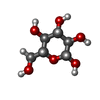
| #1: Protein | Mass: 53441.180 Da / Num. of mol.: 2 Source method: isolated from a genetically manipulated source Source: (gene. exp.)   #2: Sugar | Has ligand of interest | Y | Has protein modification | N | |
|---|
-Experimental details
-Experiment
| Experiment | Method: ELECTRON MICROSCOPY |
|---|---|
| EM experiment | Aggregation state: PARTICLE / 3D reconstruction method: single particle reconstruction |
- Sample preparation
| Component | Name: Homo-dimeric complex / Type: COMPLEX / Entity ID: #1 / Source: RECOMBINANT | ||||||||||||||||||||
|---|---|---|---|---|---|---|---|---|---|---|---|---|---|---|---|---|---|---|---|---|---|
| Molecular weight | Units: MEGADALTONS / Experimental value: NO | ||||||||||||||||||||
| Source (natural) | Organism:  | ||||||||||||||||||||
| Source (recombinant) | Organism:  | ||||||||||||||||||||
| Buffer solution | pH: 8 | ||||||||||||||||||||
| Buffer component |
| ||||||||||||||||||||
| Specimen | Conc.: 1 mg/ml / Embedding applied: NO / Shadowing applied: NO / Staining applied: NO / Vitrification applied: YES | ||||||||||||||||||||
| Specimen support | Grid material: COPPER / Grid mesh size: 200 divisions/in. / Grid type: Quantifoil R2/1 | ||||||||||||||||||||
| Vitrification | Instrument: FEI VITROBOT MARK IV / Cryogen name: ETHANE / Humidity: 100 % / Chamber temperature: 277.15 K |
- Electron microscopy imaging
| Experimental equipment |  Model: Titan Krios / Image courtesy: FEI Company |
|---|---|
| Microscopy | Model: FEI TITAN KRIOS |
| Electron gun | Electron source:  FIELD EMISSION GUN / Accelerating voltage: 300 kV / Illumination mode: FLOOD BEAM FIELD EMISSION GUN / Accelerating voltage: 300 kV / Illumination mode: FLOOD BEAM |
| Electron lens | Mode: BRIGHT FIELD / Nominal magnification: 130000 X / Nominal defocus max: 1800 nm / Nominal defocus min: 800 nm / Cs: 2.7 mm / C2 aperture diameter: 50 µm |
| Specimen holder | Cryogen: NITROGEN / Specimen holder model: FEI TITAN KRIOS AUTOGRID HOLDER |
| Image recording | Average exposure time: 1.232 sec. / Electron dose: 60.4 e/Å2 / Film or detector model: GATAN K3 BIOQUANTUM (6k x 4k) / Num. of grids imaged: 1 / Num. of real images: 12348 |
| EM imaging optics | Energyfilter name: GIF Bioquantum / Energyfilter slit width: 20 eV |
| Image scans | Width: 5760 / Height: 4092 |
- Processing
| EM software |
| ||||||||||||||||||||||||
|---|---|---|---|---|---|---|---|---|---|---|---|---|---|---|---|---|---|---|---|---|---|---|---|---|---|
| CTF correction | Type: PHASE FLIPPING AND AMPLITUDE CORRECTION | ||||||||||||||||||||||||
| Particle selection | Num. of particles selected: 4474138 | ||||||||||||||||||||||||
| Symmetry | Point symmetry: C2 (2 fold cyclic) | ||||||||||||||||||||||||
| 3D reconstruction | Resolution: 2.56 Å / Resolution method: FSC 0.143 CUT-OFF / Num. of particles: 237565 / Symmetry type: POINT | ||||||||||||||||||||||||
| Atomic model building | Protocol: OTHER / Space: REAL | ||||||||||||||||||||||||
| Atomic model building | Accession code: AF-P69786-F1 / Source name: AlphaFold / Type: in silico model |
 Movie
Movie Controller


 PDBj
PDBj


