 Movie
Movie Controller

[English] 日本語
 Yorodumi
- PDB-8qqk: Cryo-EM structure of E. coli cytochrome bo3 quinol oxidase assemb... -
+ Open data

- Basic information
| Entry | Database: PDB / ID: 8qqk | ||||||||||||
|---|---|---|---|---|---|---|---|---|---|---|---|---|---|
| Title | Cryo-EM structure of E. coli cytochrome bo3 quinol oxidase assembled in peptidiscs | ||||||||||||
 Components Components | (Cytochrome bo(3) ubiquinol oxidase subunit ...) x 4 | ||||||||||||
 Keywords Keywords | MEMBRANE PROTEIN / E. coli / Ni-NTA resin / cytochrome bo3 quinol oxidase / ubiquinone-8 release / peptidisc / single particle analysis / cryo-EM | ||||||||||||
| Function / homology |  Function and homology information Function and homology informationoxidoreduction-driven active transmembrane transporter activity / cytochrome o ubiquinol oxidase complex / ubiquinol oxidase (H+-transporting) / cytochrome bo3 ubiquinol oxidase activity / aerobic electron transport chain / respiratory chain complex / oxidoreductase activity, acting on diphenols and related substances as donors, oxygen as acceptor / cytochrome-c oxidase activity / proton transmembrane transporter activity / ubiquinone binding ...oxidoreduction-driven active transmembrane transporter activity / cytochrome o ubiquinol oxidase complex / ubiquinol oxidase (H+-transporting) / cytochrome bo3 ubiquinol oxidase activity / aerobic electron transport chain / respiratory chain complex / oxidoreductase activity, acting on diphenols and related substances as donors, oxygen as acceptor / cytochrome-c oxidase activity / proton transmembrane transporter activity / ubiquinone binding / electron transport coupled proton transport / ATP synthesis coupled electron transport / aerobic respiration / respiratory electron transport chain / electron transfer activity / copper ion binding / heme binding / plasma membrane Similarity search - Function | ||||||||||||
| Biological species |  | ||||||||||||
| Method | ELECTRON MICROSCOPY / single particle reconstruction / cryo EM / Resolution: 2.8 Å | ||||||||||||
 Authors Authors | Gao, Y. / Zhang, Y. / Hakke, S. / Peters, P.J. / Ravelli, R.B.G. | ||||||||||||
| Funding support |  Netherlands, 3items Netherlands, 3items
| ||||||||||||
 Citation Citation | Journal: Biochim Biophys Acta Bioenerg / Year: 2024 Title: Cryo-EM structure of cytochrome bo quinol oxidase assembled in peptidiscs reveals an "open" conformation for potential ubiquinone-8 release. Authors: Ye Gao / Yue Zhang / Sneha Hakke / Ronny Mohren / Lyanne J P M Sijbers / Peter J Peters / Raimond B G Ravelli / Abstract: Cytochrome bo quinol oxidase belongs to the heme‑copper-oxidoreductase (HCO) superfamily, which is part of the respiratory chain and essential for cell survival. While the reaction mechanism of cyt ...Cytochrome bo quinol oxidase belongs to the heme‑copper-oxidoreductase (HCO) superfamily, which is part of the respiratory chain and essential for cell survival. While the reaction mechanism of cyt bo has been studied extensively over the last decades, specific details about its substrate binding and product release have remained unelucidated due to the lack of structural information. Here, we report a 2.8 Å cryo-electron microscopy structure of cyt bo from Escherichia coli assembled in peptidiscs. Our structural model shows a conformation for amino acids 1-41 of subunit I different from all previously published structures while the remaining parts of this enzyme are similar. Our new conformation shows a "U-shape" assembly in contrast to the transmembrane helix, named "TM0", in other reported structural models. However, TM0 blocks ubiquinone-8 (reaction product) release, suggesting that other cyt bo conformations should exist. Our structural model presents experimental evidence for an "open" conformation to facilitate substrate/product exchange. This work helps further understand the reaction cycle of this oxidase, which could be a benefit for potential drug/antibiotic design for health science. | ||||||||||||
| History |
|
- Structure visualization
| Structure viewer | Molecule: MolmilJmol/JSmol |
|---|
- Downloads & links
-Download
| PDBx/mmCIF format | 8qqk.cif.gz | 236.6 KB | Display | PDBx/mmCIF format |
|---|---|---|---|---|
| PDB format | pdb8qqk.ent.gz | 182.5 KB | Display | PDB format |
| PDBx/mmJSON format | 8qqk.json.gz | Tree view | PDBx/mmJSON format | |
| Others |  Other downloads Other downloads |
-Validation report
| Arichive directory | https://data.pdbj.org/pub/pdb/validation_reports/qq/8qqkftp://data.pdbj.org/pub/pdb/validation_reports/qq/8qqk | HTTPS FTP |
|---|
-Related structure data
| Related structure data |  18594MC M: map data used to model this data C: citing same article ( |
|---|---|
| Similar structure data |
-Links
 PDBj
PDBj


- Assembly
| Deposited unit | 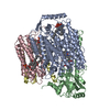
|
|---|---|
| 1 |
|
-Components
-Cytochrome bo(3) ubiquinol oxidase subunit ... , 4 types, 4 molecules ABCD
| #1: Protein | Mass: 74424.469 Da / Num. of mol.: 1 / Source method: isolated from a natural source / Source: (natural) References: UniProt: P0ABI8, ubiquinol oxidase (H+-transporting) |
|---|---|
| #2: Protein | Mass: 34947.203 Da / Num. of mol.: 1 / Source method: isolated from a natural source / Source: (natural) |
| #3: Protein | Mass: 22642.566 Da / Num. of mol.: 1 / Source method: isolated from a natural source / Source: (natural) |
| #4: Protein | Mass: 12037.402 Da / Num. of mol.: 1 / Source method: isolated from a natural source / Source: (natural) |
-Non-polymers , 5 types, 9 molecules 


| #5: Chemical |  Mass: 760.076 Da / Num. of mol.: 3 / Source method: obtained synthetically / Formula: C42H82NO8P / Comment: phospholipid*YM Mass: 760.076 Da / Num. of mol.: 3 / Source method: obtained synthetically / Formula: C42H82NO8P / Comment: phospholipid*YM#6: Chemical |  Mass: 748.065 Da / Num. of mol.: 3 / Source method: obtained synthetically / Formula: C41H82NO8P / Comment: phospholipid*YM Mass: 748.065 Da / Num. of mol.: 3 / Source method: obtained synthetically / Formula: C41H82NO8P / Comment: phospholipid*YM#7: Chemical | ChemComp-HEM / |  Mass: 616.487 Da / Num. of mol.: 1 / Source method: obtained synthetically / Formula: C34H32FeN4O4 Mass: 616.487 Da / Num. of mol.: 1 / Source method: obtained synthetically / Formula: C34H32FeN4O4#8: Chemical | ChemComp-HEO / |  Mass: 838.854 Da / Num. of mol.: 1 / Source method: obtained synthetically / Formula: C49H58FeN4O5 Mass: 838.854 Da / Num. of mol.: 1 / Source method: obtained synthetically / Formula: C49H58FeN4O5#9: Chemical | ChemComp-CU / |  Mass: 63.546 Da / Num. of mol.: 1 / Source method: obtained synthetically / Formula: Cu Mass: 63.546 Da / Num. of mol.: 1 / Source method: obtained synthetically / Formula: Cu |
|---|
-Details
| Has ligand of interest | N |
|---|
-Experimental details
-Experiment
| Experiment | Method: ELECTRON MICROSCOPY |
|---|---|
| EM experiment | Aggregation state: PARTICLE / 3D reconstruction method: single particle reconstruction |
- Sample preparation
| Component | Name: Cytochrome bo3 quinol oxidase / Type: COMPLEX Details: Heme-copper-oxidoreductase purified from E. coli native membranes by Ni-NTA affinity chromatography. Entity ID: #1-#4 / Source: NATURAL | ||||||||||||||||||||
|---|---|---|---|---|---|---|---|---|---|---|---|---|---|---|---|---|---|---|---|---|---|
| Molecular weight | Value: 0.144 MDa / Experimental value: NO | ||||||||||||||||||||
| Source (natural) | Organism: | ||||||||||||||||||||
| Buffer solution | pH: 8 Details: 20mM Tris-HCL, 150mM NaCl, 2% Glycerol, filtered and degassed. | ||||||||||||||||||||
| Buffer component |
| ||||||||||||||||||||
| Specimen | Conc.: 1.6 mg/ml / Embedding applied: NO / Shadowing applied: NO / Staining applied: NO / Vitrification applied: YES | ||||||||||||||||||||
| Specimen support | Grid material: GOLD / Grid mesh size: 300 divisions/in. / Grid type: UltrAuFoil R1.2/1.3 | ||||||||||||||||||||
| Vitrification | Instrument: FEI VITROBOT MARK IV / Cryogen name: ETHANE / Humidity: 95 % / Chamber temperature: 277.15 K |
- Electron microscopy imaging
| Experimental equipment |  Model: Titan Krios / Image courtesy: FEI Company |
|---|---|
| Microscopy | Model: FEI TITAN KRIOS |
| Electron gun | Electron source:  FIELD EMISSION GUN / Accelerating voltage: 300 kV / Illumination mode: FLOOD BEAM FIELD EMISSION GUN / Accelerating voltage: 300 kV / Illumination mode: FLOOD BEAM |
| Electron lens | Mode: BRIGHT FIELD / Nominal magnification: 105000 X / Nominal defocus max: 2000 nm / Nominal defocus min: 1500 nm / C2 aperture diameter: 100 µm / Alignment procedure: ZEMLIN TABLEAU |
| Specimen holder | Cryogen: NITROGEN / Specimen holder model: FEI TITAN KRIOS AUTOGRID HOLDER |
| Image recording | Average exposure time: 2.1 sec. / Electron dose: 50 e/Å2 / Film or detector model: GATAN K3 BIOQUANTUM (6k x 4k) / Num. of grids imaged: 1 / Num. of real images: 8050 |
| Image scans | Sampling size: 5 µm / Width: 5760 / Height: 4092 |
- Processing
| EM software |
| ||||||||||||||||||||||||||||||
|---|---|---|---|---|---|---|---|---|---|---|---|---|---|---|---|---|---|---|---|---|---|---|---|---|---|---|---|---|---|---|---|
| CTF correction | Type: PHASE FLIPPING AND AMPLITUDE CORRECTION | ||||||||||||||||||||||||||||||
| Particle selection | Num. of particles selected: 1277595 | ||||||||||||||||||||||||||||||
| Symmetry | Point symmetry: C1 (asymmetric) | ||||||||||||||||||||||||||||||
| 3D reconstruction | Resolution: 2.8 Å / Resolution method: FSC 0.143 CUT-OFF / Num. of particles: 45014 / Algorithm: FOURIER SPACE / Num. of class averages: 1 / Symmetry type: POINT | ||||||||||||||||||||||||||||||
| Atomic model building | B value: 59.97 / Protocol: FLEXIBLE FIT / Space: REAL | ||||||||||||||||||||||||||||||
| Atomic model building | PDB-ID: 1FFT Accession code: 1FFT / Source name: PDB / Type: experimental model | ||||||||||||||||||||||||||||||
| Refine LS restraints |
|

