 Movie
Movie Controller

[English] 日本語
 Yorodumi
- PDB-8qpx: Crystal structure of Geodia cydonium Immunoglobulin-like domain o... -
+ Open data

- Basic information
| Entry | Database: PDB / ID: 8qpx | |||||||||
|---|---|---|---|---|---|---|---|---|---|---|
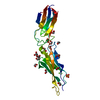
| Title | Crystal structure of Geodia cydonium Immunoglobulin-like domain of Receptor Tyrosine Kinase | |||||||||
 Components Components | Receptor tyrosine kinase (Fragment) | |||||||||
 Keywords Keywords | IMMUNE SYSTEM / Ig-like / porifera / evolution | |||||||||
| Function / homology |  Function and homology information Function and homology information | |||||||||
| Biological species |  Geodia cydonium (invertebrata) Geodia cydonium (invertebrata) | |||||||||
| Method |  X-RAY DIFFRACTION / SYNCHROTRON / MOLECULAR REPLACEMENT / Resolution: 2.1 Å X-RAY DIFFRACTION / SYNCHROTRON / MOLECULAR REPLACEMENT / Resolution: 2.1 Å | |||||||||
 Authors Authors | Lopez-Sagaseta, J. / Urdiciain, A. | |||||||||
| Funding support |  Spain, 2items Spain, 2items
| |||||||||
 Citation Citation | Journal: Commun Biol / Year: 2025 Title: Unusual traits shape the architecture of the Ig ancestor molecule. Authors: Urdiciain, A. / Madej, T. / Wang, J. / Song, J. / Erausquin, E. / Youkharibache, P. / Lopez-Sagaseta, J. #1: Journal: Sci Rep / Year: 2025 Title: Severe deviation in protein fold prediction by advanced AI: a case study. Authors: Lopez-Sagaseta, J. / Urdiciain, A. | |||||||||
| History |
|
- Structure visualization
| Structure viewer | Molecule: MolmilJmol/JSmol |
|---|
- Downloads & links
-Download
| PDBx/mmCIF format | 8qpx.cif.gz | 116.1 KB | Display | PDBx/mmCIF format |
|---|---|---|---|---|
| PDB format | pdb8qpx.ent.gz | 72.7 KB | Display | PDB format |
| PDBx/mmJSON format | 8qpx.json.gz | Tree view | PDBx/mmJSON format | |
| Others |  Other downloads Other downloads |
-Validation report
| Summary document | 8qpx_validation.pdf.gz | 869 KB | Display | wwPDB validaton report |
|---|---|---|---|---|
| Full document | 8qpx_full_validation.pdf.gz | 869.8 KB | Display | |
| Data in XML | 8qpx_validation.xml.gz | 10.9 KB | Display | |
| Data in CIF | 8qpx_validation.cif.gz | 14.8 KB | Display | |
| Arichive directory | https://data.pdbj.org/pub/pdb/validation_reports/qp/8qpxftp://data.pdbj.org/pub/pdb/validation_reports/qp/8qpx | HTTPS FTP |
-Related structure data
-Links
 PDBj
PDBj

- Assembly
| Deposited unit | 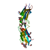
| ||||||||||||
|---|---|---|---|---|---|---|---|---|---|---|---|---|---|
| 1 |
| ||||||||||||
| Unit cell |
|
-Components
-Protein , 1 types, 1 molecules A
| #1: Protein | Mass: 24623.100 Da / Num. of mol.: 1 Source method: isolated from a genetically manipulated source Details: Remaining N-terminal GP motif from 3C site / Source: (gene. exp.) Geodia cydonium (invertebrata) / Gene: TK_2L / Production host:   Spodoptera frugiperda (fall armyworm) / References: UniProt: O18431 Spodoptera frugiperda (fall armyworm) / References: UniProt: O18431 |
|---|
-Sugars , 2 types, 2 molecules 
| #2: Polysaccharide | alpha-D-mannopyranose-(1-3)-[alpha-D-mannopyranose-(1-6)]beta-D-mannopyranose-(1-4)-2-acetamido-2- ...alpha-D-mannopyranose-(1-3)-[alpha-D-mannopyranose-(1-6)]beta-D-mannopyranose-(1-4)-2-acetamido-2-deoxy-beta-D-glucopyranose-(1-4)-[alpha-L-fucopyranose-(1-6)]2-acetamido-2-deoxy-beta-D-glucopyranose Source method: isolated from a genetically manipulated source |
|---|---|
| #3: Sugar | ChemComp-NAG /  Type: D-saccharide, beta linking / Mass: 221.208 Da / Num. of mol.: 1 / Source method: obtained synthetically / Formula: C8H15NO6 Type: D-saccharide, beta linking / Mass: 221.208 Da / Num. of mol.: 1 / Source method: obtained synthetically / Formula: C8H15NO6 |
-Non-polymers , 3 types, 105 molecules 


| #4: Chemical | ChemComp-EDO /  Mass: 62.068 Da / Num. of mol.: 5 / Source method: obtained synthetically / Formula: C2H6O2 Mass: 62.068 Da / Num. of mol.: 5 / Source method: obtained synthetically / Formula: C2H6O2#5: Chemical |  Mass: 96.063 Da / Num. of mol.: 2 / Source method: isolated from a natural source / Formula: SO4 Mass: 96.063 Da / Num. of mol.: 2 / Source method: isolated from a natural source / Formula: SO4#6: Water | ChemComp-HOH / | Mass: 18.015 Da / Num. of mol.: 98 / Source method: isolated from a natural source / Formula: H2O |
|---|
-Details
| Has ligand of interest | N |
|---|---|
| Has protein modification | Y |
-Experimental details
-Experiment
| Experiment | Method: X-RAY DIFFRACTION / Number of used crystals: 1 |
|---|
- Sample preparation
| Crystal | Density Matthews: 2.5 Å3/Da / Density % sol: 50.85 % |
|---|---|
| Crystal grow | Temperature: 295 K / Method: vapor diffusion, sitting drop / Details: 0.2 M Ammonium sulfate, 26% w/v PEG4000 |
-Data collection
| Diffraction | Mean temperature: 100 K / Serial crystal experiment: N |
|---|---|
| Diffraction source | Source: SYNCHROTRON / Site: ALBA / Beamline: XALOC / Wavelength: 0.97926 Å |
| Detector | Type: DECTRIS PILATUS 6M / Detector: PIXEL / Date: Sep 22, 2023 |
| Radiation | Monochromator: Si(111) channel cut / Protocol: SINGLE WAVELENGTH / Monochromatic (M) / Laue (L): M / Scattering type: x-ray |
| Radiation wavelength | Wavelength: 0.97926 Å / Relative weight: 1 |
| Reflection | Resolution: 2.1→40.91 Å / Num. obs: 47524 / % possible obs: 98.6 % / Redundancy: 3.3 % / Biso Wilson estimate: 34.44 Å2 / CC1/2: 0.999 / Rmerge(I) obs: 0.035 / Rpim(I) all: 0.023 / Χ2: 0.9 / Net I/σ(I): 17.4 |
| Reflection shell | Resolution: 2.1→2.16 Å / Redundancy: 3.4 % / Rmerge(I) obs: 0.247 / Mean I/σ(I) obs: 5 / Num. unique obs: 3990 / CC1/2: 0.947 / Rpim(I) all: 0.157 / Χ2: 1 / % possible all: 98.7 |
- Processing
| Software |
| ||||||||||||||||||||||||||||||||||||||||||
|---|---|---|---|---|---|---|---|---|---|---|---|---|---|---|---|---|---|---|---|---|---|---|---|---|---|---|---|---|---|---|---|---|---|---|---|---|---|---|---|---|---|---|---|
| Refinement | Method to determine structure: MOLECULAR REPLACEMENT / Resolution: 2.1→40.91 Å / SU ML: 0.2881 / Cross valid method: FREE R-VALUE / σ(F): 1.34 / Phase error: 29.6235 Stereochemistry target values: GeoStd + Monomer Library + CDL v1.2
| ||||||||||||||||||||||||||||||||||||||||||
| Solvent computation | Shrinkage radii: 0.9 Å / VDW probe radii: 1.1 Å / Solvent model: FLAT BULK SOLVENT MODEL | ||||||||||||||||||||||||||||||||||||||||||
| Displacement parameters | Biso mean: 44.41 Å2 | ||||||||||||||||||||||||||||||||||||||||||
| Refinement step | Cycle: LAST / Resolution: 2.1→40.91 Å
| ||||||||||||||||||||||||||||||||||||||||||
| Refine LS restraints |
| ||||||||||||||||||||||||||||||||||||||||||
| LS refinement shell |
| ||||||||||||||||||||||||||||||||||||||||||
| Refinement TLS params. | Method: refined / Origin x: 9.31014388062 Å / Origin y: -9.79483222786 Å / Origin z: 6.9598198259 Å
| ||||||||||||||||||||||||||||||||||||||||||
| Refinement TLS group | Selection details: (chain A and resseq 134:407) |
