 Movie
Movie Controller

[English] 日本語
 Yorodumi
- PDB-8qpm: Structure of methylene-tetrahydromethanopterin reductase from Met... -
+ Open data

- Basic information
| Entry | Database: PDB / ID: 8qpm | ||||||
|---|---|---|---|---|---|---|---|
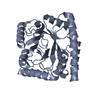
| Title | Structure of methylene-tetrahydromethanopterin reductase from Methanocaldococcus jannaschii | ||||||
 Components Components | 5,10-methylenetetrahydromethanopterin reductase | ||||||
 Keywords Keywords | OXIDOREDUCTASE / methylene-tetrahydropterin reductase | ||||||
| Function / homology |  Function and homology information Function and homology information5,10-methylenetetrahydromethanopterin reductase / coenzyme F420-dependent N5,N10-methenyltetrahydromethanopterin reductase activity / methanogenesis, from carbon dioxide / oxidoreductase activity, acting on paired donors, with incorporation or reduction of molecular oxygen / one-carbon metabolic process / cytoplasm Similarity search - Function | ||||||
| Biological species |   Methanocaldococcus jannaschii (archaea) Methanocaldococcus jannaschii (archaea) | ||||||
| Method |  X-RAY DIFFRACTION / SYNCHROTRON / MOLECULAR REPLACEMENT / Resolution: 1.8 Å X-RAY DIFFRACTION / SYNCHROTRON / MOLECULAR REPLACEMENT / Resolution: 1.8 Å | ||||||
 Authors Authors | Gehl, M. / Demmer, U. / Ermler, U. / Shima, S. | ||||||
| Funding support |  Germany, 1items Germany, 1items
| ||||||
 Citation Citation | Journal: Protein Sci. / Year: 2024 Title: Mutational and structural studies of ( beta alpha ) 8 -barrel fold methylene-tetrahydropterin reductases utilizing a common catalytic mechanism. Authors: Gehl, M. / Demmer, U. / Ermler, U. / Shima, S. | ||||||
| History |
|
- Structure visualization
| Structure viewer | Molecule: MolmilJmol/JSmol |
|---|
- Downloads & links
-Download
| PDBx/mmCIF format | 8qpm.cif.gz | 723.5 KB | Display | PDBx/mmCIF format |
|---|---|---|---|---|
| PDB format | pdb8qpm.ent.gz | 616.5 KB | Display | PDB format |
| PDBx/mmJSON format | 8qpm.json.gz | Tree view | PDBx/mmJSON format | |
| Others |  Other downloads Other downloads |
-Validation report
| Arichive directory | https://data.pdbj.org/pub/pdb/validation_reports/qp/8qpmftp://data.pdbj.org/pub/pdb/validation_reports/qp/8qpm | HTTPS FTP |
|---|
-Related structure data


-Links
 PDBj
PDBj
- Assembly
| Deposited unit | 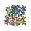
| ||||||||
|---|---|---|---|---|---|---|---|---|---|
| 1 | 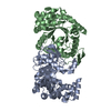
| ||||||||
| 2 | 
| ||||||||
| Unit cell |
|
-Components
| #1: Protein | Mass: 35836.656 Da / Num. of mol.: 4 Source method: isolated from a genetically manipulated source Source: (gene. exp.) Methanocaldococcus jannaschii (archaea)Gene: mer, HA335_01095 / Production host:  References: UniProt: A0A832SYB5, 5,10-methylenetetrahydromethanopterin reductase #2: Water | ChemComp-HOH / |  Mass: 18.015 Da / Num. of mol.: 657 / Source method: isolated from a natural source / Formula: H2O Mass: 18.015 Da / Num. of mol.: 657 / Source method: isolated from a natural source / Formula: H2OHas ligand of interest | N | |
|---|
-Experimental details
-Experiment
| Experiment | Method: X-RAY DIFFRACTION / Number of used crystals: 1 |
|---|
- Sample preparation
| Crystal | Density Matthews: 2.74 Å3/Da / Density % sol: 55.12 % |
|---|---|
| Crystal grow | Temperature: 281 K / Method: vapor diffusion, sitting drop Details: 35% (v/v) 2-methyl-2,4-pentanediol, 100 mM sodium acetate pH 4.5 |
-Data collection
| Diffraction | Mean temperature: 100 K / Serial crystal experiment: N |
|---|---|
| Diffraction source | Source: SYNCHROTRON / Site: SLS  / Beamline: X10SA / Wavelength: 1 Å / Beamline: X10SA / Wavelength: 1 Å |
| Detector | Type: DECTRIS EIGER2 X 16M / Detector: PIXEL / Date: Feb 10, 2021 |
| Radiation | Protocol: SINGLE WAVELENGTH / Monochromatic (M) / Laue (L): M / Scattering type: x-ray |
| Radiation wavelength | Wavelength: 1 Å / Relative weight: 1 |
| Reflection | Resolution: 1.8→48.33 Å / Num. obs: 144127 / % possible obs: 99.84 % / Redundancy: 3.8 % / CC1/2: 0.994 / Rmerge(I) obs: 0.109 / Net I/σ(I): 8.3 |
| Reflection shell | Resolution: 1.8→1.864 Å / Rmerge(I) obs: 0.822 / Num. unique obs: 14203 / CC1/2: 0.503 |
- Processing
| Software |
| ||||||||||||||||
|---|---|---|---|---|---|---|---|---|---|---|---|---|---|---|---|---|---|
| Refinement | Method to determine structure: MOLECULAR REPLACEMENT / Resolution: 1.8→48.33 Å / Cross valid method: FREE R-VALUE
| ||||||||||||||||
| Refinement step | Cycle: LAST / Resolution: 1.8→48.33 Å
|
