 Movie
Movie Controller


+ Open data

- Basic information
| Entry | Database: PDB / ID: 8qpf | |||||||||
|---|---|---|---|---|---|---|---|---|---|---|
| Title | Ammonium Transporter Amt1 from Shewanella denitrificans | |||||||||
 Components Components | Ammonium transporter | |||||||||
 Keywords Keywords | TRANSPORT PROTEIN / Ammonium Transporter Amt1 | |||||||||
| Function / homology |  Function and homology information Function and homology informationammonium homeostasis / ammonium channel activity / catalytic activity / plasma membrane Similarity search - Function | |||||||||
| Biological species |  Shewanella denitrificans (bacteria) Shewanella denitrificans (bacteria) | |||||||||
| Method | ELECTRON MICROSCOPY / single particle reconstruction / cryo EM / Resolution: 2.53 Å | |||||||||
 Authors Authors | Gschell, M. / Zhang, L. / Einsle, O. / Andrade, S. | |||||||||
| Funding support |  Germany, 2items Germany, 2items
| |||||||||
 Citation Citation | Journal: Sci Adv / Year: 2024 Title: How sensor Amt-like proteins integrate ammonium signals. Authors: Tobias Pflüger / Mathias Gschell / Lin Zhang / Volodymyr Shnitsar / Annas J Zabadné / Paul Zierep / Stefan Günther / Oliver Einsle / Susana L A Andrade / Abstract: Unlike aquaporins or potassium channels, ammonium transporters (Amts) uniquely discriminate ammonium from potassium and water. This feature has certainly contributed to their repurposing as ammonium ...Unlike aquaporins or potassium channels, ammonium transporters (Amts) uniquely discriminate ammonium from potassium and water. This feature has certainly contributed to their repurposing as ammonium receptors during evolution. Here, we describe the ammonium receptor Sd-Amt1, where an Amt module connects to a cytoplasmic diguanylate cyclase transducer module via an HAMP domain. Structures of the protein with and without bound ammonium were determined to 1.7- and 1.9-Ångstrom resolution, depicting the ON and OFF states of the receptor and confirming the presence of a binding site for two ammonium cations that is pivotal for signal perception and receptor activation. The transducer domain was disordered in the crystals, and an AlphaFold2 prediction suggests that the helices linking both domains are flexible. While the sensor domain retains the trimeric fold formed by all Amt family members, the HAMP domains interact as pairs and serve to dimerize the transducer domain upon activation. | |||||||||
| History |
|
- Structure visualization
| Structure viewer | Molecule: MolmilJmol/JSmol |
|---|
- Downloads & links
-Download
| PDBx/mmCIF format | 8qpf.cif.gz | 238.7 KB | Display | PDBx/mmCIF format |
|---|---|---|---|---|
| PDB format | pdb8qpf.ent.gz | 185.5 KB | Display | PDB format |
| PDBx/mmJSON format | 8qpf.json.gz | Tree view | PDBx/mmJSON format | |
| Others |  Other downloads Other downloads |
-Validation report
| Arichive directory | https://data.pdbj.org/pub/pdb/validation_reports/qp/8qpfftp://data.pdbj.org/pub/pdb/validation_reports/qp/8qpf | HTTPS FTP |
|---|
-Related structure data
| Related structure data |  18549MC  8qj3C  8qj4C M: map data used to model this data C: citing same article ( |
|---|---|
| Similar structure data |
-Links
 PDBj
PDBj


- Assembly
| Deposited unit | 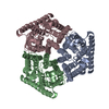
|
|---|---|
| 1 |
|
-Components
| #1: Protein | Mass: 73021.875 Da / Num. of mol.: 3 Source method: isolated from a genetically manipulated source Source: (gene. exp.) Shewanella denitrificans (bacteria) / Gene: Sden_0766 / Production host: #2: Water | ChemComp-HOH / |  Mass: 18.015 Da / Num. of mol.: 202 / Source method: isolated from a natural source / Formula: H2O Mass: 18.015 Da / Num. of mol.: 202 / Source method: isolated from a natural source / Formula: H2O |
|---|
-Experimental details
-Experiment
| Experiment | Method: ELECTRON MICROSCOPY |
|---|---|
| EM experiment | Aggregation state: PARTICLE / 3D reconstruction method: single particle reconstruction |
- Sample preparation
| Component | Name: homotrimer Amt1 / Type: COMPLEX / Entity ID: #1 / Source: RECOMBINANT |
|---|---|
| Molecular weight | Experimental value: NO |
| Source (natural) | Organism: Shewanella denitrificans (bacteria) |
| Source (recombinant) | Organism: |
| Buffer solution | pH: 7.5 |
| Specimen | Embedding applied: NO / Shadowing applied: NO / Staining applied: NO / Vitrification applied: YES |
| Vitrification | Cryogen name: ETHANE |
- Electron microscopy imaging
| Experimental equipment |  Model: Titan Krios / Image courtesy: FEI Company |
|---|---|
| Microscopy | Model: FEI TITAN KRIOS |
| Electron gun | Electron source:  FIELD EMISSION GUN / Accelerating voltage: 300 kV / Illumination mode: SPOT SCAN FIELD EMISSION GUN / Accelerating voltage: 300 kV / Illumination mode: SPOT SCAN |
| Electron lens | Mode: DARK FIELD / Nominal defocus max: 3000 nm / Nominal defocus min: 1000 nm |
| Image recording | Electron dose: 50 e/Å2 / Film or detector model: GATAN K3 (6k x 4k) |
- Processing
| EM software | Name: PHENIX / Version: 1.20.1_4487: / Category: model refinement | ||||||||||||||||||||||||
|---|---|---|---|---|---|---|---|---|---|---|---|---|---|---|---|---|---|---|---|---|---|---|---|---|---|
| CTF correction | Type: NONE | ||||||||||||||||||||||||
| 3D reconstruction | Resolution: 2.53 Å / Resolution method: FSC 0.143 CUT-OFF / Num. of particles: 477507 / Symmetry type: POINT | ||||||||||||||||||||||||
| Refine LS restraints |
|
