 Movie
Movie Controller

[English] 日本語
 Yorodumi
- PDB-8qnd: Crystal structure of the ribonucleoside hydrolase C from Lactobac... -
+ Open data

- Basic information
| Entry | Database: PDB / ID: 8qnd | ||||||
|---|---|---|---|---|---|---|---|
| Title | Crystal structure of the ribonucleoside hydrolase C from Lactobacillus reuteri | ||||||
 Components Components | Inosine-uridine nucleoside N-ribohydrolase | ||||||
 Keywords Keywords | HYDROLASE / Ribonucleoside hydrolase C / lactobacilli | ||||||
| Function / homology | : / hydrolase activity, hydrolyzing N-glycosyl compounds / Inosine/uridine-preferring nucleoside hydrolase / Inosine/uridine-preferring nucleoside hydrolase domain / Inosine-uridine preferring nucleoside hydrolase / Ribonucleoside hydrolase-like / nucleobase-containing compound metabolic process / Inosine-uridine nucleoside N-ribohydrolase Function and homology information Function and homology information | ||||||
| Biological species |  Limosilactobacillus reuteri (bacteria) Limosilactobacillus reuteri (bacteria) | ||||||
| Method |  X-RAY DIFFRACTION / MOLECULAR REPLACEMENT / Resolution: 1.9 Å X-RAY DIFFRACTION / MOLECULAR REPLACEMENT / Resolution: 1.9 Å | ||||||
 Authors Authors | Matyuta, I.O. / Minyaev, M.E. / Shaposhnikov, L.A. / Pometun, E.V. / Tishkov, V.I. / Popov, V.O. / Boyko, K.M. | ||||||
| Funding support |  Russian Federation, 1items Russian Federation, 1items
| ||||||
 Citation Citation | Journal: Int J Mol Sci / Year: 2023 Title: Structure-Functional Examination of Novel Ribonucleoside Hydrolase C (RihC) from Limosilactobacillus reuteri LR1. Authors: Shaposhnikov, L.A. / Chikurova, N.Y. / Atroshenko, D.L. / Savin, S.S. / Kleymenov, S.Y. / Chernobrovkina, A.V. / Pometun, E.V. / Minyaev, M.E. / Matyuta, I.O. / Hushpulian, D.M. / Boyko, K.M. ...Authors: Shaposhnikov, L.A. / Chikurova, N.Y. / Atroshenko, D.L. / Savin, S.S. / Kleymenov, S.Y. / Chernobrovkina, A.V. / Pometun, E.V. / Minyaev, M.E. / Matyuta, I.O. / Hushpulian, D.M. / Boyko, K.M. / Tishkov, V.I. / Pometun, A.A. | ||||||
| History |
|
- Structure visualization
| Structure viewer | Molecule: MolmilJmol/JSmol |
|---|
- Downloads & links
-Download
| PDBx/mmCIF format | 8qnd.cif.gz | 248.8 KB | Display | PDBx/mmCIF format |
|---|---|---|---|---|
| PDB format | pdb8qnd.ent.gz | 197.3 KB | Display | PDB format |
| PDBx/mmJSON format | 8qnd.json.gz | Tree view | PDBx/mmJSON format | |
| Others |  Other downloads Other downloads |
-Validation report
| Summary document | 8qnd_validation.pdf.gz | 3.1 MB | Display | wwPDB validaton report |
|---|---|---|---|---|
| Full document | 8qnd_full_validation.pdf.gz | 3.1 MB | Display | |
| Data in XML | 8qnd_validation.xml.gz | 48.1 KB | Display | |
| Data in CIF | 8qnd_validation.cif.gz | 71.3 KB | Display | |
| Arichive directory | https://data.pdbj.org/pub/pdb/validation_reports/qn/8qndftp://data.pdbj.org/pub/pdb/validation_reports/qn/8qnd | HTTPS FTP |
-Related structure data
| Similar structure data |
|---|
-Links
 PDBj
PDBj- Assembly
| Deposited unit | 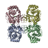
| |||||||||||||||||||||
|---|---|---|---|---|---|---|---|---|---|---|---|---|---|---|---|---|---|---|---|---|---|---|
| 1 |
| |||||||||||||||||||||
| Unit cell |
| |||||||||||||||||||||
| Noncrystallographic symmetry (NCS) | NCS domain:
|
-Components
| #1: Protein | Mass: 33419.824 Da / Num. of mol.: 4 Source method: isolated from a genetically manipulated source Source: (gene. exp.) Limosilactobacillus reuteri (bacteria) / Gene: rihC / Production host: #2: Chemical | ChemComp-CA /   Mass: 40.078 Da / Num. of mol.: 6 / Source method: obtained synthetically / Formula: Ca / Feature type: SUBJECT OF INVESTIGATION Mass: 40.078 Da / Num. of mol.: 6 / Source method: obtained synthetically / Formula: Ca / Feature type: SUBJECT OF INVESTIGATION#3: Water | ChemComp-HOH / |  Mass: 18.015 Da / Num. of mol.: 766 / Source method: isolated from a natural source / Formula: H2O Mass: 18.015 Da / Num. of mol.: 766 / Source method: isolated from a natural source / Formula: H2OHas ligand of interest | Y | |
|---|
-Experimental details
-Experiment
| Experiment | Method: X-RAY DIFFRACTION / Number of used crystals: 1 |
|---|
- Sample preparation
| Crystal | Density Matthews: 2.22 Å3/Da / Density % sol: 44.5 % |
|---|---|
| Crystal grow | Temperature: 288 K / Method: vapor diffusion, sitting drop Details: 0.2 M Magnesium acetate tetrahydrate, 0.1 M Sodium cacodylate trihydrate pH 6.5, 20% w/v Polyethylene glycol 8,000 |
-Data collection
| Diffraction | Mean temperature: 100 K / Serial crystal experiment: N |
|---|---|
| Diffraction source | Source: ROTATING ANODE / Type: RIGAKU / Wavelength: 1.5418 Å |
| Detector | Type: RIGAKU HyPix-6000HE / Detector: PIXEL / Date: Jul 18, 2022 |
| Radiation | Protocol: SINGLE WAVELENGTH / Monochromatic (M) / Laue (L): M / Scattering type: x-ray |
| Radiation wavelength | Wavelength: 1.5418 Å / Relative weight: 1 |
| Reflection | Resolution: 1.9→63.38 Å / Num. obs: 91739 / % possible obs: 99.8 % / Redundancy: 6.4 % / CC1/2: 0.914 / Rmerge(I) obs: 0.082 / Rpim(I) all: 0.039 / Rrim(I) all: 0.091 / Χ2: 1.03 / Net I/σ(I): 19.5 / Num. measured all: 585566 |
| Reflection shell | Resolution: 1.9→1.93 Å / % possible obs: 100 % / Redundancy: 6.6 % / Rmerge(I) obs: 0.347 / Num. measured all: 29786 / Num. unique obs: 4547 / CC1/2: 0.956 / Rpim(I) all: 0.146 / Rrim(I) all: 0.377 / Χ2: 1 / Net I/σ(I) obs: 5.5 |
- Processing
| Software |
| ||||||||||||||||||||||||||||||||||||||||||||||||||||||||||||||||||||||||||||||||||||||||||||||||||||||||||||||||||||||||||||||||||||||||||||||||||||||||||||||||||||||||||||||||||||||
|---|---|---|---|---|---|---|---|---|---|---|---|---|---|---|---|---|---|---|---|---|---|---|---|---|---|---|---|---|---|---|---|---|---|---|---|---|---|---|---|---|---|---|---|---|---|---|---|---|---|---|---|---|---|---|---|---|---|---|---|---|---|---|---|---|---|---|---|---|---|---|---|---|---|---|---|---|---|---|---|---|---|---|---|---|---|---|---|---|---|---|---|---|---|---|---|---|---|---|---|---|---|---|---|---|---|---|---|---|---|---|---|---|---|---|---|---|---|---|---|---|---|---|---|---|---|---|---|---|---|---|---|---|---|---|---|---|---|---|---|---|---|---|---|---|---|---|---|---|---|---|---|---|---|---|---|---|---|---|---|---|---|---|---|---|---|---|---|---|---|---|---|---|---|---|---|---|---|---|---|---|---|---|---|
| Refinement | Method to determine structure: MOLECULAR REPLACEMENT / Resolution: 1.9→63.38 Å / Cor.coef. Fo:Fc: 0.954 / Cor.coef. Fo:Fc free: 0.938 / SU B: 2.871 / SU ML: 0.085 / Cross valid method: THROUGHOUT / ESU R: 0.145 / ESU R Free: 0.129 / Stereochemistry target values: MAXIMUM LIKELIHOOD / Details: HYDROGENS HAVE BEEN ADDED IN THE RIDING POSITIONS
| ||||||||||||||||||||||||||||||||||||||||||||||||||||||||||||||||||||||||||||||||||||||||||||||||||||||||||||||||||||||||||||||||||||||||||||||||||||||||||||||||||||||||||||||||||||||
| Solvent computation | Ion probe radii: 0.8 Å / Shrinkage radii: 0.8 Å / VDW probe radii: 1.2 Å / Solvent model: MASK | ||||||||||||||||||||||||||||||||||||||||||||||||||||||||||||||||||||||||||||||||||||||||||||||||||||||||||||||||||||||||||||||||||||||||||||||||||||||||||||||||||||||||||||||||||||||
| Displacement parameters | Biso mean: 22.401 Å2
| ||||||||||||||||||||||||||||||||||||||||||||||||||||||||||||||||||||||||||||||||||||||||||||||||||||||||||||||||||||||||||||||||||||||||||||||||||||||||||||||||||||||||||||||||||||||
| Refinement step | Cycle: 1 / Resolution: 1.9→63.38 Å
| ||||||||||||||||||||||||||||||||||||||||||||||||||||||||||||||||||||||||||||||||||||||||||||||||||||||||||||||||||||||||||||||||||||||||||||||||||||||||||||||||||||||||||||||||||||||
| Refine LS restraints |
|