 Movie
Movie Controller

[English] 日本語
 Yorodumi
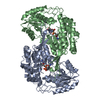
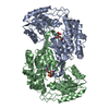
- PDB-8qmt: Succinic semialdehyde dehydrogenase from E. coli with Q262R subst... -
+ Open data

- Basic information
| Entry | Database: PDB / ID: 8qmt | ||||||
|---|---|---|---|---|---|---|---|
| Title | Succinic semialdehyde dehydrogenase from E. coli with Q262R substitution and bound NAD+, succinic semialdehyde | ||||||
 Components Components | Succinate semialdehyde dehydrogenase [NAD(P)+] Sad | ||||||
 Keywords Keywords | OXIDOREDUCTASE / succinic semialdehyde / dehydrogenase | ||||||
| Function / homology |  Function and homology information Function and homology informationsuccinate-semialdehyde dehydrogenase [NAD(P)+] activity / succinate-semialdehyde dehydrogenase (NADP+) activity / succinate-semialdehyde dehydrogenase [NAD(P)+] / putrescine catabolic process / succinate-semialdehyde dehydrogenase (NAD+) activity / cellular detoxification of nitrogen compound / GABA catabolic process / aldehyde dehydrogenase [NAD(P)+] activity / L-arginine catabolic process / tricarboxylic acid cycle / protein homodimerization activity Similarity search - Function | ||||||
| Biological species |  | ||||||
| Method |  X-RAY DIFFRACTION / SYNCHROTRON / MOLECULAR REPLACEMENT / Resolution: 1.8 Å X-RAY DIFFRACTION / SYNCHROTRON / MOLECULAR REPLACEMENT / Resolution: 1.8 Å | ||||||
 Authors Authors | He, H. / Zarzycki, J. / Erb, T.J. | ||||||
| Funding support |  Germany, 1items Germany, 1items
| ||||||
 Citation Citation | Journal: Nat Commun / Year: 2024 Title: Adaptive laboratory evolution recruits the promiscuity of succinate semialdehyde dehydrogenase to repair different metabolic deficiencies. Authors: He, H. / Gomez-Coronado, P.A. / Zarzycki, J. / Barthel, S. / Kahnt, J. / Claus, P. / Klein, M. / Klose, M. / de Crecy-Lagard, V. / Schindler, D. / Paczia, N. / Glatter, T. / Erb, T.J. | ||||||
| History |
|
- Structure visualization
| Structure viewer | Molecule: MolmilJmol/JSmol |
|---|
- Downloads & links
-Download
| PDBx/mmCIF format | 8qmt.cif.gz | 715.1 KB | Display | PDBx/mmCIF format |
|---|---|---|---|---|
| PDB format | pdb8qmt.ent.gz | 594.4 KB | Display | PDB format |
| PDBx/mmJSON format | 8qmt.json.gz | Tree view | PDBx/mmJSON format | |
| Others |  Other downloads Other downloads |
-Validation report
| Arichive directory | https://data.pdbj.org/pub/pdb/validation_reports/qm/8qmtftp://data.pdbj.org/pub/pdb/validation_reports/qm/8qmt | HTTPS FTP |
|---|
-Related structure data
-Links
 PDBj
PDBj

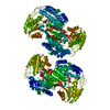
- Assembly
| Deposited unit | 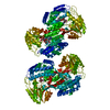
| ||||||||
|---|---|---|---|---|---|---|---|---|---|
| 1 | 
| ||||||||
| 2 | 
| ||||||||
| Unit cell |
|
-Components
| #1: Protein | Mass: 49798.754 Da / Num. of mol.: 4 / Mutation: Q262R Source method: isolated from a genetically manipulated source Source: (gene. exp.) References: UniProt: P76149, succinate-semialdehyde dehydrogenase [NAD(P)+] #2: Chemical | ChemComp-NAD /   Mass: 663.425 Da / Num. of mol.: 4 / Source method: obtained synthetically / Formula: C21H27N7O14P2 / Feature type: SUBJECT OF INVESTIGATION / Comment: NAD*YM Mass: 663.425 Da / Num. of mol.: 4 / Source method: obtained synthetically / Formula: C21H27N7O14P2 / Feature type: SUBJECT OF INVESTIGATION / Comment: NAD*YM#3: Chemical | ChemComp-SSN / 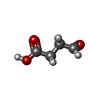  Mass: 102.089 Da / Num. of mol.: 4 / Source method: obtained synthetically / Formula: C4H6O3 / Feature type: SUBJECT OF INVESTIGATION Mass: 102.089 Da / Num. of mol.: 4 / Source method: obtained synthetically / Formula: C4H6O3 / Feature type: SUBJECT OF INVESTIGATION#4: Water | ChemComp-HOH / |  Mass: 18.015 Da / Num. of mol.: 1293 / Source method: isolated from a natural source / Formula: H2O Mass: 18.015 Da / Num. of mol.: 1293 / Source method: isolated from a natural source / Formula: H2OHas ligand of interest | Y | Has protein modification | Y | |
|---|
-Experimental details
-Experiment
| Experiment | Method: X-RAY DIFFRACTION / Number of used crystals: 1 |
|---|
- Sample preparation
| Crystal | Density Matthews: 2.38 Å3/Da / Density % sol: 48.25 % |
|---|---|
| Crystal grow | Temperature: 295 K / Method: vapor diffusion, sitting drop / pH: 7.5 Details: 14.8 mg/mL of SAD_Q262R in 20 mM HEPES-KOH, 50 mM KCl, pH 7.5 was complemented with 5 mM NAD+. The enzyme was then mixed in a 2:1 ratio with 250 mM Bis-Tris-Propane, pH 7.5 , 20 % (w/v) ...Details: 14.8 mg/mL of SAD_Q262R in 20 mM HEPES-KOH, 50 mM KCl, pH 7.5 was complemented with 5 mM NAD+. The enzyme was then mixed in a 2:1 ratio with 250 mM Bis-Tris-Propane, pH 7.5 , 20 % (w/v) PEG4000. The final drop size was 3 uL. The drop was complemented with 6 mM succininc semialdehyde and 25 % (v/v) PEG400 before flash freezing the crystals in liquid nitrogen. |
-Data collection
| Diffraction | Mean temperature: 100 K / Serial crystal experiment: N | ||||||||||||||||||||||||||||||||||||||||||||||||||||||||||||||||||||||||||||||||||||||||||||||||||||||||||||||||||||||||||||||
|---|---|---|---|---|---|---|---|---|---|---|---|---|---|---|---|---|---|---|---|---|---|---|---|---|---|---|---|---|---|---|---|---|---|---|---|---|---|---|---|---|---|---|---|---|---|---|---|---|---|---|---|---|---|---|---|---|---|---|---|---|---|---|---|---|---|---|---|---|---|---|---|---|---|---|---|---|---|---|---|---|---|---|---|---|---|---|---|---|---|---|---|---|---|---|---|---|---|---|---|---|---|---|---|---|---|---|---|---|---|---|---|---|---|---|---|---|---|---|---|---|---|---|---|---|---|---|---|
| Diffraction source | Source: SYNCHROTRON / Site: PETRA III, EMBL c/o DESY / Beamline: P13 (MX1) / Wavelength: 0.9763 Å | ||||||||||||||||||||||||||||||||||||||||||||||||||||||||||||||||||||||||||||||||||||||||||||||||||||||||||||||||||||||||||||||
| Detector | Type: DECTRIS EIGER X 16M / Detector: PIXEL / Date: Jun 20, 2023 | ||||||||||||||||||||||||||||||||||||||||||||||||||||||||||||||||||||||||||||||||||||||||||||||||||||||||||||||||||||||||||||||
| Radiation | Protocol: SINGLE WAVELENGTH / Monochromatic (M) / Laue (L): M / Scattering type: x-ray | ||||||||||||||||||||||||||||||||||||||||||||||||||||||||||||||||||||||||||||||||||||||||||||||||||||||||||||||||||||||||||||||
| Radiation wavelength | Wavelength: 0.9763 Å / Relative weight: 1 | ||||||||||||||||||||||||||||||||||||||||||||||||||||||||||||||||||||||||||||||||||||||||||||||||||||||||||||||||||||||||||||||
| Reflection | Resolution: 1.8→29.55 Å / Num. obs: 174478 / % possible obs: 98.6 % / Redundancy: 13.8 % / CC1/2: 0.999 / Rmerge(I) obs: 0.098 / Rrim(I) all: 0.102 / Net I/σ(I): 16.73 | ||||||||||||||||||||||||||||||||||||||||||||||||||||||||||||||||||||||||||||||||||||||||||||||||||||||||||||||||||||||||||||||
| Reflection shell |
|
- Processing
| Software |
| |||||||||||||||||||||||||||||||||||||||||||||||||||||||||||||||||||||||||||||||||||||||||||||||||||||||||
|---|---|---|---|---|---|---|---|---|---|---|---|---|---|---|---|---|---|---|---|---|---|---|---|---|---|---|---|---|---|---|---|---|---|---|---|---|---|---|---|---|---|---|---|---|---|---|---|---|---|---|---|---|---|---|---|---|---|---|---|---|---|---|---|---|---|---|---|---|---|---|---|---|---|---|---|---|---|---|---|---|---|---|---|---|---|---|---|---|---|---|---|---|---|---|---|---|---|---|---|---|---|---|---|---|---|---|
| Refinement | Method to determine structure: MOLECULAR REPLACEMENT / Resolution: 1.8→29.55 Å / SU ML: 0.21 / Cross valid method: FREE R-VALUE / σ(F): 1.34 / Phase error: 24.57 / Stereochemistry target values: ML
| |||||||||||||||||||||||||||||||||||||||||||||||||||||||||||||||||||||||||||||||||||||||||||||||||||||||||
| Solvent computation | Shrinkage radii: 0.9 Å / VDW probe radii: 1.1 Å / Solvent model: FLAT BULK SOLVENT MODEL | |||||||||||||||||||||||||||||||||||||||||||||||||||||||||||||||||||||||||||||||||||||||||||||||||||||||||
| Refinement step | Cycle: LAST / Resolution: 1.8→29.55 Å
| |||||||||||||||||||||||||||||||||||||||||||||||||||||||||||||||||||||||||||||||||||||||||||||||||||||||||
| Refine LS restraints |
| |||||||||||||||||||||||||||||||||||||||||||||||||||||||||||||||||||||||||||||||||||||||||||||||||||||||||
| LS refinement shell |
| |||||||||||||||||||||||||||||||||||||||||||||||||||||||||||||||||||||||||||||||||||||||||||||||||||||||||
| Refinement TLS params. | Method: refined / Origin x: -22.4993 Å / Origin y: -29.453 Å / Origin z: -30.2199 Å
| |||||||||||||||||||||||||||||||||||||||||||||||||||||||||||||||||||||||||||||||||||||||||||||||||||||||||
| Refinement TLS group | Selection details: all |
