[English] 日本語
 Yorodumi
- PDB-8qmp: Structure of the E2 Beryllium Fluoride Complex of the Autoinhibit... -
+ Open data
- Basic information
| Entry | Database: PDB / ID: 8qmp | ||||||
|---|---|---|---|---|---|---|---|
| Title | Structure of the E2 Beryllium Fluoride Complex of the Autoinhibited Calcium ATPase ACA8 | ||||||
 Components Components | Calcium-transporting ATPase 8, plasma membrane-type | ||||||
 Keywords Keywords | TRANSPORT PROTEIN / HYDROLASE Calcium transporter P-type ATPase / HYDROLASE | ||||||
| Function / homology |  Function and homology information Function and homology informationP-type Ca2+ transporter / P-type calcium transporter activity / plasmodesma / response to nematode / plastid / calmodulin binding / ATP hydrolysis activity / ATP binding / metal ion binding / plasma membrane Similarity search - Function | ||||||
| Biological species |  | ||||||
| Method | ELECTRON MICROSCOPY / single particle reconstruction / cryo EM / Resolution: 3.3 Å | ||||||
 Authors Authors | Thirup Larsen, S. / Karlsen Dannersoe, J. / Nissen, P. | ||||||
| Funding support |  Denmark, 1items Denmark, 1items
| ||||||
 Citation Citation |  Journal: J Mol Biol / Year: 2024 Journal: J Mol Biol / Year: 2024Title: Conserved N-terminal Regulation of the ACA8 Calcium Pump with Two Calmodulin Binding Sites. Authors: Sigrid Thirup Larsen / Josephine Karlsen Dannersø / Christine Juul Fælled Nielsen / Lisbeth Rosager Poulsen / Michael Palmgren / Poul Nissen /  Abstract: The autoinhibited plasma membrane calcium ATPase ACA8 from A. thaliana has an N-terminal autoinhibitory domain. Binding of calcium-loaded calmodulin at two sites located at residues 42-62 and 74-96 ...The autoinhibited plasma membrane calcium ATPase ACA8 from A. thaliana has an N-terminal autoinhibitory domain. Binding of calcium-loaded calmodulin at two sites located at residues 42-62 and 74-96 relieves autoinhibition of ACA8 activity. Through activity studies and a yeast complementation assay we investigated wild-type (WT) and N-terminally truncated ACA8 constructs (Δ20, Δ30, Δ35, Δ37, Δ40, Δ74 and Δ100) to explore the role of conserved motifs in the N-terminal segment preceding the calmodulin binding sites. Furthermore, we purified WT, Δ20- and Δ100-ACA8, tested activity in vitro and performed structural studies of purified Δ20-ACA8 stabilized in a lipid nanodisc to explore the mechanism of autoinhibition. We show that an N-terminal segment between residues 20 and 35 including conserved Phe32, upstream of the calmodulin binding sites, is important for autoinhibition and the activation by calmodulin. Cryo-EM structure determination at 3.3 Å resolution of a beryllium fluoride inhibited E2 form, and at low resolution for an E1 state combined with AlphaFold prediction provide a model for autoinhibition, consistent with the mutational studies. | ||||||
| History |
|
- Structure visualization
| Structure viewer | Molecule:  Molmil Molmil Jmol/JSmol Jmol/JSmol |
|---|
- Downloads & links
- Download
| PDBx/mmCIF format |  8qmp.cif.gz 8qmp.cif.gz | 342.3 KB | Display |  PDBx/mmCIF format PDBx/mmCIF format |
|---|---|---|---|---|
| PDB format |  pdb8qmp.ent.gz pdb8qmp.ent.gz | 277.2 KB | Display |  PDB format PDB format |
| PDBx/mmJSON format |  8qmp.json.gz 8qmp.json.gz | Tree view |  PDBx/mmJSON format PDBx/mmJSON format | |
| Others |  Other downloads Other downloads |
-Validation report
| Summary document |  8qmp_validation.pdf.gz 8qmp_validation.pdf.gz | 1.3 MB | Display |  wwPDB validaton report wwPDB validaton report |
|---|---|---|---|---|
| Full document |  8qmp_full_validation.pdf.gz 8qmp_full_validation.pdf.gz | 1.3 MB | Display | |
| Data in XML |  8qmp_validation.xml.gz 8qmp_validation.xml.gz | 39.9 KB | Display | |
| Data in CIF |  8qmp_validation.cif.gz 8qmp_validation.cif.gz | 58.6 KB | Display | |
| Arichive directory |  https://data.pdbj.org/pub/pdb/validation_reports/qm/8qmp https://data.pdbj.org/pub/pdb/validation_reports/qm/8qmp ftp://data.pdbj.org/pub/pdb/validation_reports/qm/8qmp ftp://data.pdbj.org/pub/pdb/validation_reports/qm/8qmp | HTTPS FTP |
-Related structure data
| Related structure data |  18506MC M: map data used to model this data C: citing same article ( |
|---|---|
| Similar structure data | Similarity search - Function & homology  F&H Search F&H Search |
- Links
Links
- Assembly
| Deposited unit | 
|
|---|---|
| 1 |
|
- Components
| #1: Protein | Mass: 114233.523 Da / Num. of mol.: 1 Source method: isolated from a genetically manipulated source Source: (gene. exp.)   |
|---|---|
| #2: Chemical | ChemComp-MG / |
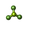
| #3: Chemical | ChemComp-BEF / |
| Has ligand of interest | N |
| Has protein modification | Y |
-Experimental details
-Experiment
| Experiment | Method: ELECTRON MICROSCOPY |
|---|---|
| EM experiment | Aggregation state: PARTICLE / 3D reconstruction method: single particle reconstruction |
- Sample preparation
| Component | Name: ACA8 beryllium fluoride complex / Type: COMPLEX / Entity ID: #1 / Source: RECOMBINANT | ||||||||||||||||||||||||||||||
|---|---|---|---|---|---|---|---|---|---|---|---|---|---|---|---|---|---|---|---|---|---|---|---|---|---|---|---|---|---|---|---|
| Molecular weight | Value: 0.116 MDa / Experimental value: NO | ||||||||||||||||||||||||||||||
| Source (natural) | Organism:  | ||||||||||||||||||||||||||||||
| Source (recombinant) | Organism:  | ||||||||||||||||||||||||||||||
| Buffer solution | pH: 7.5 | ||||||||||||||||||||||||||||||
| Buffer component |
| ||||||||||||||||||||||||||||||
| Specimen | Conc.: 0.67 mg/ml / Embedding applied: NO / Shadowing applied: NO / Staining applied: NO / Vitrification applied: YES | ||||||||||||||||||||||||||||||
| Specimen support | Grid material: COPPER / Grid mesh size: 300 divisions/in. / Grid type: C-flat-1.2/1.3 | ||||||||||||||||||||||||||||||
| Vitrification | Instrument: LEICA PLUNGER / Cryogen name: ETHANE / Humidity: 90 % / Chamber temperature: 283 K |
- Electron microscopy imaging
| Experimental equipment |  Model: Titan Krios / Image courtesy: FEI Company |
|---|---|
| Microscopy | Model: FEI TITAN KRIOS |
| Electron gun | Electron source:  FIELD EMISSION GUN / Accelerating voltage: 300 kV / Illumination mode: FLOOD BEAM FIELD EMISSION GUN / Accelerating voltage: 300 kV / Illumination mode: FLOOD BEAM |
| Electron lens | Mode: BRIGHT FIELD / Nominal magnification: 130000 X / Nominal defocus max: 1800 nm / Nominal defocus min: 800 nm |
| Image recording | Electron dose: 60 e/Å2 / Film or detector model: GATAN K3 (6k x 4k) |
- Processing
| EM software |
| ||||||||||||||||||||||||||||||||||||
|---|---|---|---|---|---|---|---|---|---|---|---|---|---|---|---|---|---|---|---|---|---|---|---|---|---|---|---|---|---|---|---|---|---|---|---|---|---|
| CTF correction | Type: PHASE FLIPPING AND AMPLITUDE CORRECTION | ||||||||||||||||||||||||||||||||||||
| Particle selection | Num. of particles selected: 941952 | ||||||||||||||||||||||||||||||||||||
| 3D reconstruction | Resolution: 3.3 Å / Resolution method: FSC 0.143 CUT-OFF / Num. of particles: 193419 / Symmetry type: POINT | ||||||||||||||||||||||||||||||||||||
| Refine LS restraints |
|
 Movie
Movie Controller


 PDBj
PDBj


