 Movie
Movie Controller

[English] 日本語
 Yorodumi
- PDB-8qk1: Crystal structure of Trichuris suis beta-N-acetyl-D-hexosaminidas... -
+ Open data

- Basic information
| Entry | Database: PDB / ID: 8qk1 | ||||||
|---|---|---|---|---|---|---|---|
| Title | Crystal structure of Trichuris suis beta-N-acetyl-D-hexosaminidase - HEX-2 in apo form | ||||||
 Components Components | beta-N-acetylhexosaminidase | ||||||
 Keywords Keywords | HYDROLASE / hexosaminidase / enzyme / Glycoside Hydrolase Family 20 | ||||||
| Function / homology |  Function and homology information Function and homology information: / beta-N-acetylhexosaminidase activity / beta-N-acetylhexosaminidase / carbohydrate metabolic process / membrane Similarity search - Function | ||||||
| Biological species |  Trichuris suis (pig whipworm) Trichuris suis (pig whipworm) | ||||||
| Method |  X-RAY DIFFRACTION / SYNCHROTRON / MOLECULAR REPLACEMENT / Resolution: 2.55 Å X-RAY DIFFRACTION / SYNCHROTRON / MOLECULAR REPLACEMENT / Resolution: 2.55 Å | ||||||
 Authors Authors | Dutkiewicz, Z. / Varrot, A. | ||||||
| Funding support |  Austria, 1items Austria, 1items
| ||||||
 Citation Citation | Journal: Biochemistry / Year: 2024 Title: Bioinformatic, Enzymatic, and Structural Characterization of Trichuris suis Hexosaminidase HEX-2. Authors: Dutkiewicz, Z. / Varrot, A. / Breese, K.J. / Stubbs, K.A. / Nuschy, L. / Adduci, I. / Paschinger, K. / Wilson, I.B.H. | ||||||
| History |
|
- Structure visualization
| Structure viewer | Molecule: MolmilJmol/JSmol |
|---|
- Downloads & links
-Download
| PDBx/mmCIF format | 8qk1.cif.gz | 112.4 KB | Display | PDBx/mmCIF format |
|---|---|---|---|---|
| PDB format | pdb8qk1.ent.gz | 80.6 KB | Display | PDB format |
| PDBx/mmJSON format | 8qk1.json.gz | Tree view | PDBx/mmJSON format | |
| Others |  Other downloads Other downloads |
-Validation report
| Arichive directory | https://data.pdbj.org/pub/pdb/validation_reports/qk/8qk1ftp://data.pdbj.org/pub/pdb/validation_reports/qk/8qk1 | HTTPS FTP |
|---|
-Related structure data
| Similar structure data | |
|---|---|
| Experimental dataset #1 | Data reference: 10.5281/zenodo.8367522 / Data set type: diffraction image data / Metadata reference: 10.5281/zenodo.8367522 |
-Links
 PDBj
PDBj- Assembly
| Deposited unit | 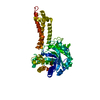
| ||||||||
|---|---|---|---|---|---|---|---|---|---|
| 1 |
| ||||||||
| Unit cell |
|
-Components
| #1: Protein | Mass: 63269.191 Da / Num. of mol.: 1 Source method: isolated from a genetically manipulated source Source: (gene. exp.) Trichuris suis (pig whipworm) / Gene: M514_07548 / Production host:  Komagataella pastoris (fungus) / Strain (production host): GS115 / References: UniProt: A0A085NCI8 Komagataella pastoris (fungus) / Strain (production host): GS115 / References: UniProt: A0A085NCI8 |
|---|---|
| #2: Chemical | ChemComp-ZN /   Mass: 65.409 Da / Num. of mol.: 1 / Source method: obtained synthetically / Formula: Zn / Feature type: SUBJECT OF INVESTIGATION Mass: 65.409 Da / Num. of mol.: 1 / Source method: obtained synthetically / Formula: Zn / Feature type: SUBJECT OF INVESTIGATION |
| #3: Water | ChemComp-HOH /  Mass: 18.015 Da / Num. of mol.: 44 / Source method: isolated from a natural source / Formula: H2O Mass: 18.015 Da / Num. of mol.: 44 / Source method: isolated from a natural source / Formula: H2O |
| Has ligand of interest | Y |
| Has protein modification | Y |
-Experimental details
-Experiment
| Experiment | Method: X-RAY DIFFRACTION / Number of used crystals: 1 |
|---|
- Sample preparation
| Crystal | Density Matthews: 2.17 Å3/Da / Density % sol: 43.2 % |
|---|---|
| Crystal grow | Temperature: 292 K / Method: vapor diffusion, hanging drop / pH: 7.5 Details: 20% PEG 1500 0.15M potasium thiocynate 0.1M Tris pH 7.5 |
-Data collection
| Diffraction | Mean temperature: 100 K / Serial crystal experiment: N |
|---|---|
| Diffraction source | Source: SYNCHROTRON / Site: SOLEIL  / Beamline: PROXIMA 1 / Wavelength: 0.97856 Å / Beamline: PROXIMA 1 / Wavelength: 0.97856 Å |
| Detector | Type: DECTRIS EIGER X 16M / Detector: PIXEL / Date: Sep 10, 2022 |
| Radiation | Protocol: SINGLE WAVELENGTH / Monochromatic (M) / Laue (L): M / Scattering type: x-ray |
| Radiation wavelength | Wavelength: 0.97856 Å / Relative weight: 1 |
| Reflection | Resolution: 2.55→43.74 Å / Num. obs: 17835 / % possible obs: 99.9 % / Redundancy: 6.5 % / Biso Wilson estimate: 59.7 Å2 / CC1/2: 0.999 / Rmerge(I) obs: 0.067 / Rpim(I) all: 0.043 / Rrim(I) all: 0.08 / Net I/σ(I): 14.1 |
| Reflection shell | Resolution: 2.55→2.66 Å / Redundancy: 6.7 % / Rmerge(I) obs: 0.782 / Mean I/σ(I) obs: 2.1 / Num. unique obs: 2157 / CC1/2: 0.843 / Rpim(I) all: 0.5 / Rrim(I) all: 0.931 / % possible all: 99.9 |
- Processing
| Software |
| |||||||||||||||||||||||||||||||||||||||||||||||||||||||||||||||||||||||||||||||||||||||||||||||||||||||||||||||||||||||||||||||||||||||||||||||||||||||||||
|---|---|---|---|---|---|---|---|---|---|---|---|---|---|---|---|---|---|---|---|---|---|---|---|---|---|---|---|---|---|---|---|---|---|---|---|---|---|---|---|---|---|---|---|---|---|---|---|---|---|---|---|---|---|---|---|---|---|---|---|---|---|---|---|---|---|---|---|---|---|---|---|---|---|---|---|---|---|---|---|---|---|---|---|---|---|---|---|---|---|---|---|---|---|---|---|---|---|---|---|---|---|---|---|---|---|---|---|---|---|---|---|---|---|---|---|---|---|---|---|---|---|---|---|---|---|---|---|---|---|---|---|---|---|---|---|---|---|---|---|---|---|---|---|---|---|---|---|---|---|---|---|---|---|---|---|---|
| Refinement | Method to determine structure: MOLECULAR REPLACEMENT / Resolution: 2.55→43.74 Å / Cor.coef. Fo:Fc: 0.949 / Cor.coef. Fo:Fc free: 0.907 / SU B: 12.767 / SU ML: 0.269 / Cross valid method: FREE R-VALUE / ESU R: 0.785 / ESU R Free: 0.324 Details: Hydrogens have been added in their riding positions
| |||||||||||||||||||||||||||||||||||||||||||||||||||||||||||||||||||||||||||||||||||||||||||||||||||||||||||||||||||||||||||||||||||||||||||||||||||||||||||
| Solvent computation | Ion probe radii: 0.8 Å / Shrinkage radii: 0.8 Å / VDW probe radii: 1.2 Å / Solvent model: MASK BULK SOLVENT | |||||||||||||||||||||||||||||||||||||||||||||||||||||||||||||||||||||||||||||||||||||||||||||||||||||||||||||||||||||||||||||||||||||||||||||||||||||||||||
| Displacement parameters | Biso mean: 42.658 Å2
| |||||||||||||||||||||||||||||||||||||||||||||||||||||||||||||||||||||||||||||||||||||||||||||||||||||||||||||||||||||||||||||||||||||||||||||||||||||||||||
| Refinement step | Cycle: LAST / Resolution: 2.55→43.74 Å
| |||||||||||||||||||||||||||||||||||||||||||||||||||||||||||||||||||||||||||||||||||||||||||||||||||||||||||||||||||||||||||||||||||||||||||||||||||||||||||
| Refine LS restraints |
| |||||||||||||||||||||||||||||||||||||||||||||||||||||||||||||||||||||||||||||||||||||||||||||||||||||||||||||||||||||||||||||||||||||||||||||||||||||||||||
| LS refinement shell |
|
