[English] 日本語
 Yorodumi
- PDB-8qjy: Human Adenovirus type 11 fiber knob in complex with two copies of... -
+ Open data
- Basic information
| Entry | Database: PDB / ID: 8qjy | ||||||
|---|---|---|---|---|---|---|---|
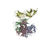
| Title | Human Adenovirus type 11 fiber knob in complex with two copies of its cell receptor, Desmoglein-2 | ||||||
 Components Components |
| ||||||
 Keywords Keywords | VIRAL PROTEIN / adenovirus / cell entry / receptor binding | ||||||
| Function / homology |  Function and homology information Function and homology informationPurkinje myocyte development / positive regulation of protein localization to cell-cell junction / bundle of His cell-Purkinje myocyte adhesion involved in cell communication / cell adhesive protein binding involved in bundle of His cell-Purkinje myocyte communication / desmosome organization / negative regulation of endothelial cell differentiation / Keratinization / negative regulation of inflammatory response to wounding / desmosome / mesenchymal to epithelial transition ...Purkinje myocyte development / positive regulation of protein localization to cell-cell junction / bundle of His cell-Purkinje myocyte adhesion involved in cell communication / cell adhesive protein binding involved in bundle of His cell-Purkinje myocyte communication / desmosome organization / negative regulation of endothelial cell differentiation / Keratinization / negative regulation of inflammatory response to wounding / desmosome / mesenchymal to epithelial transition / Formation of the cornified envelope / cornified envelope / regulation of ventricular cardiac muscle cell action potential / Apoptotic cleavage of cell adhesion proteins / adhesion receptor-mediated virion attachment to host cell / negative regulation of epithelial to mesenchymal transition / positive regulation of sprouting angiogenesis / homophilic cell-cell adhesion / positive regulation of stem cell population maintenance / regulation of heart rate by cardiac conduction / intercalated disc / lateral plasma membrane / RHOG GTPase cycle / RAC2 GTPase cycle / maternal process involved in female pregnancy / RAC3 GTPase cycle / cell adhesion molecule binding / positive regulation of cell adhesion / response to progesterone / stem cell proliferation / cell-cell adhesion / cell-cell junction / cell junction / viral capsid / cell adhesion / apical plasma membrane / intracellular membrane-bounded organelle / calcium ion binding / symbiont entry into host cell / negative regulation of apoptotic process / host cell nucleus / cell surface / extracellular exosome / plasma membrane / cytoplasm Similarity search - Function | ||||||
| Biological species |  Homo sapiens (human) Homo sapiens (human) Human adenovirus 11 Human adenovirus 11 | ||||||
| Method | ELECTRON MICROSCOPY / single particle reconstruction / cryo EM / Resolution: 3.5 Å | ||||||
 Authors Authors | Effantin, G. | ||||||
| Funding support |  France, 1items France, 1items
| ||||||
 Citation Citation |  Journal: J Virol / Year: 2023 Journal: J Virol / Year: 2023Title: Toward the understanding of DSG2 and CD46 interaction with HAdV-11 fiber, a super-complex analysis. Authors: Gregory Effantin / Marc-André Hograindleur / Daphna Fenel / Pascal Fender / Emilie Vassal-Stermann /  Abstract: The main limitation of oncolytic vectors is neutralization by blood components, which prevents intratumoral administration to patients. Enadenotucirev, a chimeric HAdV-11p/HAdV-3 adenovirus ...The main limitation of oncolytic vectors is neutralization by blood components, which prevents intratumoral administration to patients. Enadenotucirev, a chimeric HAdV-11p/HAdV-3 adenovirus identified by bio-selection, is a low seroprevalence vector active against a broad range of human carcinoma cell lines. At this stage, there's still some uncertainty about tropism and primary receptor utilization by HAdV-11. However, this information is very important, as it has a direct influence on the effectiveness of HAdV-11-based vectors. The aim of this work is to determine which of the two receptors, DSG2 and CD46, is involved in the attachment of the virus to the host, and what role they play in the early stages of infection. | ||||||
| History |
|
- Structure visualization
| Structure viewer | Molecule:  Molmil Molmil Jmol/JSmol Jmol/JSmol |
|---|
- Downloads & links
- Download
| PDBx/mmCIF format |  8qjy.cif.gz 8qjy.cif.gz | 171.2 KB | Display |  PDBx/mmCIF format PDBx/mmCIF format |
|---|---|---|---|---|
| PDB format |  pdb8qjy.ent.gz pdb8qjy.ent.gz | 121.2 KB | Display |  PDB format PDB format |
| PDBx/mmJSON format |  8qjy.json.gz 8qjy.json.gz | Tree view |  PDBx/mmJSON format PDBx/mmJSON format | |
| Others |  Other downloads Other downloads |
-Validation report
| Summary document |  8qjy_validation.pdf.gz 8qjy_validation.pdf.gz | 1.1 MB | Display |  wwPDB validaton report wwPDB validaton report |
|---|---|---|---|---|
| Full document |  8qjy_full_validation.pdf.gz 8qjy_full_validation.pdf.gz | 1.1 MB | Display | |
| Data in XML |  8qjy_validation.xml.gz 8qjy_validation.xml.gz | 32.7 KB | Display | |
| Data in CIF |  8qjy_validation.cif.gz 8qjy_validation.cif.gz | 48.5 KB | Display | |
| Arichive directory |  https://data.pdbj.org/pub/pdb/validation_reports/qj/8qjy https://data.pdbj.org/pub/pdb/validation_reports/qj/8qjy ftp://data.pdbj.org/pub/pdb/validation_reports/qj/8qjy ftp://data.pdbj.org/pub/pdb/validation_reports/qj/8qjy | HTTPS FTP |
-Related structure data
| Related structure data |  18454MC  8qjxC  8qk3C C: citing same article ( M: map data used to model this data |
|---|---|
| Similar structure data | Similarity search - Function & homology  F&H Search F&H Search |
- Links
Links
- Assembly
| Deposited unit | 
|
|---|---|
| 1 |
|
- Components
| #1: Protein | Mass: 122421.633 Da / Num. of mol.: 1 Source method: isolated from a genetically manipulated source Source: (gene. exp.)  Homo sapiens (human) / Gene: DSG2 / Production host: Homo sapiens (human) / Gene: DSG2 / Production host:  |
|---|---|
| #2: Protein | Mass: 35564.773 Da / Num. of mol.: 3 Source method: isolated from a genetically manipulated source Source: (gene. exp.)  Human adenovirus 11 / Gene: L5 / Production host: Human adenovirus 11 / Gene: L5 / Production host:  |
-Experimental details
-Experiment
| Experiment | Method: ELECTRON MICROSCOPY |
|---|---|
| EM experiment | Aggregation state: PARTICLE / 3D reconstruction method: single particle reconstruction |
- Sample preparation
| Component | Name: Complex between the human adenovirus 11 fiber knob and 1 copy of the human desmoglein 2 (domains ec2/ec3) Type: COMPLEX / Entity ID: all / Source: RECOMBINANT |
|---|---|
| Molecular weight | Experimental value: NO |
| Source (natural) | Organism:  |
| Source (recombinant) | Organism:  |
| Buffer solution | pH: 8 |
| Specimen | Embedding applied: NO / Shadowing applied: NO / Staining applied: NO / Vitrification applied: YES |
| Vitrification | Cryogen name: ETHANE |
- Electron microscopy imaging
| Experimental equipment |  Model: Titan Krios / Image courtesy: FEI Company |
|---|---|
| Microscopy | Model: FEI TITAN KRIOS |
| Electron gun | Electron source:  FIELD EMISSION GUN / Accelerating voltage: 300 kV / Illumination mode: FLOOD BEAM FIELD EMISSION GUN / Accelerating voltage: 300 kV / Illumination mode: FLOOD BEAM |
| Electron lens | Mode: BRIGHT FIELD / Nominal defocus max: 2000 nm / Nominal defocus min: 1000 nm |
| Image recording | Electron dose: 55 e/Å2 / Film or detector model: GATAN K2 SUMMIT (4k x 4k) |
- Processing
| CTF correction | Type: PHASE FLIPPING AND AMPLITUDE CORRECTION |
|---|---|
| 3D reconstruction | Resolution: 3.5 Å / Resolution method: FSC 0.143 CUT-OFF / Num. of particles: 105169 / Symmetry type: POINT |
 Movie
Movie Controller


 PDBj
PDBj


