 Movie
Movie Controller


+ Open data

- Basic information
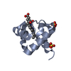
| Entry | Database: PDB / ID: 8qj6 | ||||||
|---|---|---|---|---|---|---|---|
| Title | Crystal structure of cytochrome domain 1 from PgcA | ||||||
 Components Components | Lipoprotein cytochrome c | ||||||
 Keywords Keywords | ELECTRON TRANSPORT / Geobacter sulfurreducens / cytochrome / extracellular electron transfer / mineral reduction. | ||||||
| Function / homology |  Function and homology information Function and homology informationelectron transfer activity / heme binding / metal ion binding / membrane Similarity search - Function | ||||||
| Biological species |  Geobacter sulfurreducens PCA (bacteria) Geobacter sulfurreducens PCA (bacteria) | ||||||
| Method |  X-RAY DIFFRACTION / SYNCHROTRON / MOLECULAR REPLACEMENT / Resolution: 1.55 Å X-RAY DIFFRACTION / SYNCHROTRON / MOLECULAR REPLACEMENT / Resolution: 1.55 Å | ||||||
 Authors Authors | Nash, B.W. / Edwards, M.J. / Clarke, T.A. | ||||||
| Funding support |  United Kingdom, 1items United Kingdom, 1items
| ||||||
 Citation Citation | Journal: Protein Sci. / Year: 2024 Title: Tethered heme domains in a triheme cytochrome allow for increased electron transport distances. Authors: Nash, B.W. / Fernandes, T.M. / Burton, J.A.J. / Morgado, L. / van Wonderen, J.H. / Svistunenko, D.A. / Edwards, M.J. / Salgueiro, C.A. / Butt, J.N. / Clarke, T.A. | ||||||
| History |
|
- Structure visualization
| Structure viewer | Molecule: MolmilJmol/JSmol |
|---|
- Downloads & links
-Download
| PDBx/mmCIF format | 8qj6.cif.gz | 30.6 KB | Display | PDBx/mmCIF format |
|---|---|---|---|---|
| PDB format | pdb8qj6.ent.gz | 16.8 KB | Display | PDB format |
| PDBx/mmJSON format | 8qj6.json.gz | Tree view | PDBx/mmJSON format | |
| Others |  Other downloads Other downloads |
-Validation report
| Summary document | 8qj6_validation.pdf.gz | 848.7 KB | Display | wwPDB validaton report |
|---|---|---|---|---|
| Full document | 8qj6_full_validation.pdf.gz | 848.6 KB | Display | |
| Data in XML | 8qj6_validation.xml.gz | 6.5 KB | Display | |
| Data in CIF | 8qj6_validation.cif.gz | 8 KB | Display | |
| Arichive directory | https://data.pdbj.org/pub/pdb/validation_reports/qj/8qj6ftp://data.pdbj.org/pub/pdb/validation_reports/qj/8qj6 | HTTPS FTP |
-Related structure data

-Links
 PDBj
PDBj


- Assembly
| Deposited unit | 
| ||||||||
|---|---|---|---|---|---|---|---|---|---|
| 1 |
| ||||||||
| Unit cell |
|
-Components
| #1: Protein | Mass: 9742.112 Da / Num. of mol.: 1 Source method: isolated from a genetically manipulated source Source: (gene. exp.) Geobacter sulfurreducens PCA (bacteria)Strain: PCA / Gene: pgcA, GSU1761 / Production host: Shewanella oneidensis MR-1 (bacteria) / Strain (production host): LS527 / References: UniProt: Q74CB3 |
|---|---|
| #2: Chemical | ChemComp-NA /   Mass: 22.990 Da / Num. of mol.: 1 / Source method: obtained synthetically / Formula: Na Mass: 22.990 Da / Num. of mol.: 1 / Source method: obtained synthetically / Formula: Na |
| #3: Chemical | ChemComp-HEC /   Mass: 618.503 Da / Num. of mol.: 1 / Source method: obtained synthetically / Formula: C34H34FeN4O4 / Feature type: SUBJECT OF INVESTIGATION Mass: 618.503 Da / Num. of mol.: 1 / Source method: obtained synthetically / Formula: C34H34FeN4O4 / Feature type: SUBJECT OF INVESTIGATION |
| #4: Water | ChemComp-HOH /  Mass: 18.015 Da / Num. of mol.: 69 / Source method: isolated from a natural source / Formula: H2O Mass: 18.015 Da / Num. of mol.: 69 / Source method: isolated from a natural source / Formula: H2O |
| Has ligand of interest | Y |
| Has protein modification | Y |
-Experimental details
-Experiment
| Experiment | Method: X-RAY DIFFRACTION / Number of used crystals: 1 |
|---|
- Sample preparation
| Crystal | Density Matthews: 1.66 Å3/Da / Density % sol: 26.04 % / Description: Plates |
|---|---|
| Crystal grow | Temperature: 289 K / Method: vapor diffusion, sitting drop / pH: 7 Details: 0.3 uL protein at 10 mg per mL in 20 mM HEPES pH 7.8 100 mM NaCl, 0.28 uL Na Malonate pH 7.0 and 0.02 uL 30% W/V PEG 2000 MME |
-Data collection
| Diffraction | Mean temperature: 100 K / Serial crystal experiment: N |
|---|---|
| Diffraction source | Source: SYNCHROTRON / Site: Diamond / Beamline: I04 / Wavelength: 0.9537 Å |
| Detector | Type: DECTRIS EIGER2 XE 16M / Detector: PIXEL / Date: Jul 26, 2023 |
| Radiation | Protocol: SINGLE WAVELENGTH / Monochromatic (M) / Laue (L): M / Scattering type: x-ray |
| Radiation wavelength | Wavelength: 0.9537 Å / Relative weight: 1 |
| Reflection | Resolution: 1.55→34.21 Å / Num. obs: 9230 / % possible obs: 97.4 % / Redundancy: 12.9 % / CC1/2: 0.998 / Rmerge(I) obs: 0.07 / Rpim(I) all: 0.02 / Rrim(I) all: 0.073 / Χ2: 0.96 / Net I/σ(I): 21.9 / Num. measured all: 118718 |
| Reflection shell | Resolution: 1.55→1.58 Å / % possible obs: 76.8 % / Redundancy: 8.6 % / Rmerge(I) obs: 0.512 / Num. measured all: 2980 / Num. unique obs: 345 / CC1/2: 0.915 / Rpim(I) all: 0.173 / Rrim(I) all: 0.543 / Χ2: 0.95 / Net I/σ(I) obs: 3.8 |
- Processing
| Software |
| ||||||||||||||||||||||||||||
|---|---|---|---|---|---|---|---|---|---|---|---|---|---|---|---|---|---|---|---|---|---|---|---|---|---|---|---|---|---|
| Refinement | Method to determine structure: MOLECULAR REPLACEMENT / Resolution: 1.55→34.21 Å / SU ML: 0.17 / Cross valid method: FREE R-VALUE / σ(F): 1.36 / Phase error: 19.03 / Stereochemistry target values: ML
| ||||||||||||||||||||||||||||
| Solvent computation | Shrinkage radii: 0.9 Å / VDW probe radii: 1.1 Å / Solvent model: FLAT BULK SOLVENT MODEL | ||||||||||||||||||||||||||||
| Refinement step | Cycle: LAST / Resolution: 1.55→34.21 Å
| ||||||||||||||||||||||||||||
| Refine LS restraints |
| ||||||||||||||||||||||||||||
| LS refinement shell |
|
