 Movie
Movie Controller


+ Open data

- Basic information
| Entry | Database: PDB / ID: 8qfc | |||||||||||||||||||||||||||||||||||||||||||||||||||||||||||||||||||||||||||||||||||||||||||||
|---|---|---|---|---|---|---|---|---|---|---|---|---|---|---|---|---|---|---|---|---|---|---|---|---|---|---|---|---|---|---|---|---|---|---|---|---|---|---|---|---|---|---|---|---|---|---|---|---|---|---|---|---|---|---|---|---|---|---|---|---|---|---|---|---|---|---|---|---|---|---|---|---|---|---|---|---|---|---|---|---|---|---|---|---|---|---|---|---|---|---|---|---|---|---|
| Title | UFL1 E3 ligase bound 60S ribosome | |||||||||||||||||||||||||||||||||||||||||||||||||||||||||||||||||||||||||||||||||||||||||||||
 Components Components |
| |||||||||||||||||||||||||||||||||||||||||||||||||||||||||||||||||||||||||||||||||||||||||||||
 Keywords Keywords | LIGASE / UFM1 / Ribosome / Complex | |||||||||||||||||||||||||||||||||||||||||||||||||||||||||||||||||||||||||||||||||||||||||||||
| Function / homology |  Function and homology information Function and homology informationpositive regulation of I-kappaB phosphorylation / UFM1 ligase activity / UFM1-modified protein reader activity / positive regulation of reticulophagy / regulation of phosphatase activity / apoptotic nuclear changes / definitive erythrocyte differentiation / UFM1 transferase activity / negative regulation of protein serine/threonine kinase activity / positive regulation of protein localization to endoplasmic reticulum ...positive regulation of I-kappaB phosphorylation / UFM1 ligase activity / UFM1-modified protein reader activity / positive regulation of reticulophagy / regulation of phosphatase activity / apoptotic nuclear changes / definitive erythrocyte differentiation / UFM1 transferase activity / negative regulation of protein serine/threonine kinase activity / positive regulation of protein localization to endoplasmic reticulum / protein K69-linked ufmylation / negative regulation of protein kinase activity by regulation of protein phosphorylation / protein ufmylation / : / positive regulation of plasma cell differentiation / negative regulation of IRE1-mediated unfolded protein response / regulation of proteasomal ubiquitin-dependent protein catabolic process / positive regulation of cell cycle G1/S phase transition / protein localization to endoplasmic reticulum / negative regulation of T cell mediated immune response to tumor cell / regulation of intracellular estrogen receptor signaling pathway / regulation of cyclin-dependent protein serine/threonine kinase activity / mitotic G2/M transition checkpoint / cartilage development / Transferases; Acyltransferases; Aminoacyltransferases / positive regulation of proteasomal protein catabolic process / mitogen-activated protein kinase binding / ribosome disassembly / regulation of canonical NF-kappaB signal transduction / : / response to L-glutamate / reticulophagy / regulation of neuron differentiation / negative regulation of MAP kinase activity / negative regulation of T cell activation / negative regulation of PERK-mediated unfolded protein response / negative regulation of protein phosphorylation / mitotic G2 DNA damage checkpoint signaling / ubiquitin-like protein ligase binding / RHOA GTPase cycle / positive regulation of glial cell proliferation / Peptide chain elongation / Selenocysteine synthesis / Formation of a pool of free 40S subunits / hematopoietic stem cell differentiation / Eukaryotic Translation Termination / SRP-dependent cotranslational protein targeting to membrane / Response of EIF2AK4 (GCN2) to amino acid deficiency / Viral mRNA Translation / NF-kappaB binding / Nonsense Mediated Decay (NMD) independent of the Exon Junction Complex (EJC) / positive regulation of signal transduction by p53 class mediator / GTP hydrolysis and joining of the 60S ribosomal subunit / L13a-mediated translational silencing of Ceruloplasmin expression / Major pathway of rRNA processing in the nucleolus and cytosol / ubiquitin-like ligase-substrate adaptor activity / Nonsense Mediated Decay (NMD) enhanced by the Exon Junction Complex (EJC) / endoplasmic reticulum unfolded protein response / endomembrane system / negative regulation of protein ubiquitination / MDM2/MDM4 family protein binding / negative regulation of proteasomal ubiquitin-dependent protein catabolic process / positive regulation of autophagy / regulation of mitotic cell cycle / rescue of stalled cytosolic ribosome / DNA damage checkpoint signaling / response to endoplasmic reticulum stress / cyclin binding / cytosolic ribosome / : / positive regulation of protein ubiquitination / erythrocyte differentiation / liver development / regulation of protein stability / negative regulation of protein catabolic process / brain development / positive regulation of protein localization to nucleus / Regulation of expression of SLITs and ROBOs / osteoblast differentiation / regulation of protein localization / positive regulation of proteasomal ubiquitin-dependent protein catabolic process / Antigen processing: Ubiquitination & Proteasome degradation / site of double-strand break / regulation of inflammatory response / cytosolic large ribosomal subunit / response to ethanol / cytoplasmic translation / positive regulation of canonical NF-kappaB signal transduction / mitochondrial outer membrane / cell population proliferation / neuron projection / postsynaptic density / protein stabilization / positive regulation of cell migration / structural constituent of ribosome / translation / negative regulation of gene expression / focal adhesion / DNA repair / positive regulation of cell population proliferation Similarity search - Function | |||||||||||||||||||||||||||||||||||||||||||||||||||||||||||||||||||||||||||||||||||||||||||||
| Biological species |  Homo sapiens (human) Homo sapiens (human) | |||||||||||||||||||||||||||||||||||||||||||||||||||||||||||||||||||||||||||||||||||||||||||||
| Method | ELECTRON MICROSCOPY / single particle reconstruction / cryo EM / Resolution: 3.2 Å | |||||||||||||||||||||||||||||||||||||||||||||||||||||||||||||||||||||||||||||||||||||||||||||
 Authors Authors | Makhlouf, L. / Zeqiraj, E. / Kulathu, Y. | |||||||||||||||||||||||||||||||||||||||||||||||||||||||||||||||||||||||||||||||||||||||||||||
| Funding support |  United Kingdom, European Union, 4items United Kingdom, European Union, 4items
| |||||||||||||||||||||||||||||||||||||||||||||||||||||||||||||||||||||||||||||||||||||||||||||
 Citation Citation | Journal: Nature / Year: 2024 Title: The UFM1 E3 ligase recognizes and releases 60S ribosomes from ER translocons. Authors: Linda Makhlouf / Joshua J Peter / Helge M Magnussen / Rohan Thakur / David Millrine / Thomas C Minshull / Grace Harrison / Joby Varghese / Frederic Lamoliatte / Martina Foglizzo / Thomas ...Authors: Linda Makhlouf / Joshua J Peter / Helge M Magnussen / Rohan Thakur / David Millrine / Thomas C Minshull / Grace Harrison / Joby Varghese / Frederic Lamoliatte / Martina Foglizzo / Thomas Macartney / Antonio N Calabrese / Elton Zeqiraj / Yogesh Kulathu / Abstract: Stalled ribosomes at the endoplasmic reticulum (ER) are covalently modified with the ubiquitin-like protein UFM1 on the 60S ribosomal subunit protein RPL26 (also known as uL24). This modification, ...Stalled ribosomes at the endoplasmic reticulum (ER) are covalently modified with the ubiquitin-like protein UFM1 on the 60S ribosomal subunit protein RPL26 (also known as uL24). This modification, which is known as UFMylation, is orchestrated by the UFM1 ribosome E3 ligase (UREL) complex, comprising UFL1, UFBP1 and CDK5RAP3 (ref. ). However, the catalytic mechanism of UREL and the functional consequences of UFMylation are unclear. Here we present cryo-electron microscopy structures of UREL bound to 60S ribosomes, revealing the basis of its substrate specificity. UREL wraps around the 60S subunit to form a C-shaped clamp architecture that blocks the tRNA-binding sites at one end, and the peptide exit tunnel at the other. A UFL1 loop inserts into and remodels the peptidyl transferase centre. These features of UREL suggest a crucial function for UFMylation in the release and recycling of stalled or terminated ribosomes from the ER membrane. In the absence of functional UREL, 60S-SEC61 translocon complexes accumulate at the ER membrane, demonstrating that UFMylation is necessary for releasing SEC61 from 60S subunits. Notably, this release is facilitated by a functional switch of UREL from a 'writer' to a 'reader' module that recognizes its product-UFMylated 60S ribosomes. Collectively, we identify a fundamental role for UREL in dissociating 60S subunits from the SEC61 translocon and the basis for UFMylation in regulating protein homeostasis at the ER. | |||||||||||||||||||||||||||||||||||||||||||||||||||||||||||||||||||||||||||||||||||||||||||||
| History |
|
- Structure visualization
| Structure viewer | Molecule: MolmilJmol/JSmol |
|---|
- Downloads & links
-Download
| PDBx/mmCIF format | 8qfc.cif.gz | 302.5 KB | Display | PDBx/mmCIF format |
|---|---|---|---|---|
| PDB format | pdb8qfc.ent.gz | 227.8 KB | Display | PDB format |
| PDBx/mmJSON format | 8qfc.json.gz | Tree view | PDBx/mmJSON format | |
| Others |  Other downloads Other downloads |
-Validation report
| Arichive directory | https://data.pdbj.org/pub/pdb/validation_reports/qf/8qfcftp://data.pdbj.org/pub/pdb/validation_reports/qf/8qfc | HTTPS FTP |
|---|
-Related structure data
| Related structure data |  18381MC 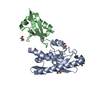 8bzrC  8c0dC  8qfdC M: map data used to model this data C: citing same article ( |
|---|---|
| Similar structure data |
-Links
 PDBj
PDBj


- Assembly
| Deposited unit | 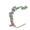
|
|---|---|
| 1 |
|
-Components
| #1: Protein | Mass: 24879.422 Da / Num. of mol.: 1 Source method: isolated from a genetically manipulated source Source: (gene. exp.) Homo sapiens (human) / Gene: RPL10A, NEDD6 / Production host:  |
|---|---|
| #2: Protein | Mass: 91591.234 Da / Num. of mol.: 1 Source method: isolated from a genetically manipulated source Source: (gene. exp.) Homo sapiens (human) / Gene: UFL1, KIAA0776, MAXER, NLBP, RCAD / Production host: References: UniProt: O94874, Transferases; Acyltransferases; Aminoacyltransferases |
| #3: Protein | Mass: 57458.938 Da / Num. of mol.: 1 Source method: isolated from a genetically manipulated source Source: (gene. exp.) Homo sapiens (human) / Gene: CDK5RAP3, IC53, LZAP, MSTP016, OK/SW-cl.114, PP1553 / Production host: |
| #4: Protein | Mass: 34644.445 Da / Num. of mol.: 1 Source method: isolated from a genetically manipulated source Source: (gene. exp.) Homo sapiens (human) / Gene: DDRGK1, C20orf116, UFBP1 / Production host: |
| #5: Protein | Mass: 9236.628 Da / Num. of mol.: 1 Source method: isolated from a genetically manipulated source Source: (gene. exp.) Homo sapiens (human) / Gene: UFM1 / Production host: |
| Has protein modification | N |
-Experimental details
-Experiment
| Experiment | Method: ELECTRON MICROSCOPY |
|---|---|
| EM experiment | Aggregation state: PARTICLE / 3D reconstruction method: single particle reconstruction |
- Sample preparation
| Component | Name: UFM1 ribosome E3 ligase complex bound to 60S ribosome / Type: COMPLEX / Entity ID: all / Source: RECOMBINANT |
|---|---|
| Source (natural) | Organism: Homo sapiens (human) |
| Source (recombinant) | Organism: |
| Buffer solution | pH: 7.5 Details: 25 mM HEPES pH 7.5, 50 mM KCl, 5 mM MgCl2, 2 mM DTT |
| Specimen | Conc.: 7.7 mg/ml / Embedding applied: NO / Shadowing applied: NO / Staining applied: NO / Vitrification applied: YES |
| Vitrification | Instrument: FEI VITROBOT MARK IV / Cryogen name: ETHANE / Humidity: 100 % / Chamber temperature: 277 K |
- Electron microscopy imaging
| Experimental equipment |  Model: Titan Krios / Image courtesy: FEI Company |
|---|---|
| Microscopy | Model: FEI TITAN KRIOS |
| Electron gun | Electron source:  FIELD EMISSION GUN / Accelerating voltage: 300 kV / Illumination mode: FLOOD BEAM FIELD EMISSION GUN / Accelerating voltage: 300 kV / Illumination mode: FLOOD BEAM |
| Electron lens | Mode: BRIGHT FIELD / Nominal magnification: 165000 X / Nominal defocus max: 2000 nm / Nominal defocus min: 200 nm |
| Image recording | Average exposure time: 2.67 sec. / Electron dose: 33.4 e/Å2 / Film or detector model: FEI FALCON IV (4k x 4k) / Num. of grids imaged: 1 / Num. of real images: 59394 |
| EM imaging optics | Energyfilter name: TFS Selectris X / Energyfilter slit width: 10 eV |
- Processing
| EM software |
| ||||||||||||||||||||||||
|---|---|---|---|---|---|---|---|---|---|---|---|---|---|---|---|---|---|---|---|---|---|---|---|---|---|
| CTF correction | Type: PHASE FLIPPING AND AMPLITUDE CORRECTION | ||||||||||||||||||||||||
| 3D reconstruction | Resolution: 3.2 Å / Resolution method: FSC 0.143 CUT-OFF / Num. of particles: 299008 / Symmetry type: POINT | ||||||||||||||||||||||||
| Refine LS restraints |
|
