 Movie
Movie Controller

[English] 日本語
 Yorodumi
- PDB-8qby: Respiratory complex I from Paracoccus denitrificans in MSP2N2 nan... -
+ Open data

- Basic information
| Entry | Database: PDB / ID: 8qby | |||||||||
|---|---|---|---|---|---|---|---|---|---|---|
| Title | Respiratory complex I from Paracoccus denitrificans in MSP2N2 nanodiscs | |||||||||
 Components Components |
| |||||||||
 Keywords Keywords | OXIDOREDUCTASE / Respiratory complex I / NADH:ubiquinone oxidoreductase / Nanodiscs | |||||||||
| Function / homology |  Function and homology information Function and homology informationactive transmembrane transporter activity / organelle envelope / protein-L-isoaspartate (D-aspartate) O-methyltransferase activity / NADH:ubiquinone reductase (H+-translocating) / : / membrane protein complex / NADH dehydrogenase (quinone) (non-electrogenic) activity / Translocases; Catalysing the translocation of protons; Linked to oxidoreductase reactions / NADH dehydrogenase complex / monoatomic cation transmembrane transporter activity ...active transmembrane transporter activity / organelle envelope / protein-L-isoaspartate (D-aspartate) O-methyltransferase activity / NADH:ubiquinone reductase (H+-translocating) / : / membrane protein complex / NADH dehydrogenase (quinone) (non-electrogenic) activity / Translocases; Catalysing the translocation of protons; Linked to oxidoreductase reactions / NADH dehydrogenase complex / monoatomic cation transmembrane transporter activity / oxidoreductase activity, acting on NAD(P)H, quinone or similar compound as acceptor / : / organelle membrane / ubiquinone binding / electron transport coupled proton transport / oxidoreductase activity, acting on NAD(P)H / NADH dehydrogenase activity / respiratory chain complex I / NADH dehydrogenase (ubiquinone) activity / quinone binding / catalytic complex / ATP synthesis coupled electron transport / endomembrane system / aerobic respiration / electron transport chain / 2 iron, 2 sulfur cluster binding / NAD binding / FMN binding / 4 iron, 4 sulfur cluster binding / response to oxidative stress / methylation / oxidoreductase activity / iron ion binding / membrane / metal ion binding / plasma membrane / cytoplasm Similarity search - Function | |||||||||
| Biological species |  Paracoccus denitrificans PD1222 (bacteria) Paracoccus denitrificans PD1222 (bacteria) | |||||||||
| Method | ELECTRON MICROSCOPY / single particle reconstruction / cryo EM / Resolution: 2.3 Å | |||||||||
 Authors Authors | Ivanov, B.S. / Bridges, H.R. / Hirst, J. | |||||||||
| Funding support |  United Kingdom, 2items United Kingdom, 2items
| |||||||||
 Citation Citation | Journal: Nat Commun / Year: 2024 Title: Structure of the turnover-ready state of an ancestral respiratory complex I. Authors: Bozhidar S Ivanov / Hannah R Bridges / Owen D Jarman / Judy Hirst /   Abstract: Respiratory complex I is pivotal for cellular energy conversion, harnessing energy from NADH:ubiquinone oxidoreduction to drive protons across energy-transducing membranes for ATP synthesis. Despite ...Respiratory complex I is pivotal for cellular energy conversion, harnessing energy from NADH:ubiquinone oxidoreduction to drive protons across energy-transducing membranes for ATP synthesis. Despite detailed structural information on complex I, its mechanism of catalysis remains elusive due to lack of accompanying functional data for comprehensive structure-function analyses. Here, we present the 2.3-Å resolution structure of complex I from the α-proteobacterium Paracoccus denitrificans, a close relative of the mitochondrial progenitor, in phospholipid-bilayer nanodiscs. Three eukaryotic-type supernumerary subunits (NDUFS4, NDUFS6 and NDUFA12) plus a novel L-isoaspartyl-O-methyltransferase are bound to the core complex. Importantly, the enzyme is in a single, homogeneous resting state that matches the closed, turnover-ready (active) state of mammalian complex I. Our structure reveals the elements that stabilise the closed state and completes P. denitrificans complex I as a unified platform for combining structure, function and genetics in mechanistic studies. | |||||||||
| History |
|
- Structure visualization
| Structure viewer | Molecule: MolmilJmol/JSmol |
|---|
- Downloads & links
-Download
| PDBx/mmCIF format | 8qby.cif.gz | 1.1 MB | Display | PDBx/mmCIF format |
|---|---|---|---|---|
| PDB format | pdb8qby.ent.gz | 857.6 KB | Display | PDB format |
| PDBx/mmJSON format | 8qby.json.gz | Tree view | PDBx/mmJSON format | |
| Others |  Other downloads Other downloads |
-Validation report
| Arichive directory | https://data.pdbj.org/pub/pdb/validation_reports/qb/8qbyftp://data.pdbj.org/pub/pdb/validation_reports/qb/8qby | HTTPS FTP |
|---|
-Related structure data
| Related structure data | 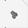 18324MC 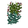 8qc1C M: map data used to model this data C: citing same article ( |
|---|---|
| Similar structure data |


-Links
 PDBj
PDBj


- Assembly
| Deposited unit | 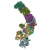
|
|---|---|
| 1 |
|
-Components
-NADH-quinone ... , 11 types, 11 molecules KGINHFDAJCB
| #1: Protein | Mass: 10863.054 Da / Num. of mol.: 1 / Source method: isolated from a natural source Source: (natural) Paracoccus denitrificans PD1222 (bacteria)References: UniProt: A1B482, Translocases; Catalysing the translocation of protons; Linked to oxidoreductase reactions |
|---|---|
| #2: Protein | Mass: 73289.398 Da / Num. of mol.: 1 / Source method: isolated from a natural source Source: (natural) Paracoccus denitrificans PD1222 (bacteria)References: UniProt: A1B489, Translocases; Catalysing the translocation of protons; Linked to oxidoreductase reactions |
| #4: Protein | Mass: 18925.561 Da / Num. of mol.: 1 / Source method: isolated from a natural source Source: (natural) Paracoccus denitrificans PD1222 (bacteria)References: UniProt: A1B486 |
| #6: Protein | Mass: 52564.820 Da / Num. of mol.: 1 / Source method: isolated from a natural source Source: (natural) Paracoccus denitrificans PD1222 (bacteria)References: UniProt: A1B479 |
| #7: Protein | Mass: 38861.152 Da / Num. of mol.: 1 / Source method: isolated from a natural source Source: (natural) Paracoccus denitrificans PD1222 (bacteria)References: UniProt: A1B487 |
| #8: Protein | Mass: 47281.141 Da / Num. of mol.: 1 / Source method: isolated from a natural source Source: (natural) Paracoccus denitrificans PD1222 (bacteria)References: UniProt: A1B491 |
| #9: Protein | Mass: 46811.484 Da / Num. of mol.: 1 / Source method: isolated from a natural source Source: (natural) Paracoccus denitrificans PD1222 (bacteria)References: UniProt: A1B495 |
| #11: Protein | Mass: 13686.093 Da / Num. of mol.: 1 / Source method: isolated from a natural source Source: (natural) Paracoccus denitrificans PD1222 (bacteria)References: UniProt: A1B498 |
| #12: Protein | Mass: 21835.205 Da / Num. of mol.: 1 / Source method: isolated from a natural source Source: (natural) Paracoccus denitrificans PD1222 (bacteria)References: UniProt: P29922 |
| #15: Protein | Mass: 23920.102 Da / Num. of mol.: 1 / Source method: isolated from a natural source Source: (natural) Paracoccus denitrificans PD1222 (bacteria)References: UniProt: A1B496 |
| #16: Protein | Mass: 19525.543 Da / Num. of mol.: 1 / Source method: isolated from a natural source Source: (natural) Paracoccus denitrificans PD1222 (bacteria)References: UniProt: A1B497 |
-Protein , 4 types, 4 molecules tqRQ
| #3: Protein | Mass: 23528.947 Da / Num. of mol.: 1 / Source method: isolated from a natural source Source: (natural) Paracoccus denitrificans PD1222 (bacteria)References: UniProt: A1B5L6 |
|---|---|
| #10: Protein | Mass: 14432.875 Da / Num. of mol.: 1 / Source method: isolated from a natural source Source: (natural) Paracoccus denitrificans PD1222 (bacteria)References: UniProt: A1B1H8 |
| #13: Protein | Mass: 7069.931 Da / Num. of mol.: 1 / Source method: isolated from a natural source Source: (natural) Paracoccus denitrificans PD1222 (bacteria)References: UniProt: A1B357 |
| #14: Protein | Mass: 12048.399 Da / Num. of mol.: 1 / Source method: isolated from a natural source Source: (natural) Paracoccus denitrificans PD1222 (bacteria)References: UniProt: A1B1M0 |
-NADH dehydrogenase subunit ... , 3 types, 3 molecules ELM
| #5: Protein | Mass: 26145.865 Da / Num. of mol.: 1 / Source method: isolated from a natural source Source: (natural) Paracoccus denitrificans PD1222 (bacteria)References: UniProt: A1B494 |
|---|---|
| #17: Protein | Mass: 77811.352 Da / Num. of mol.: 1 / Source method: isolated from a natural source Source: (natural) Paracoccus denitrificans PD1222 (bacteria)References: UniProt: A1B481 |
| #18: Protein | Mass: 56519.906 Da / Num. of mol.: 1 / Source method: isolated from a natural source Source: (natural) Paracoccus denitrificans PD1222 (bacteria)References: UniProt: A1B480 |
-Non-polymers , 13 types, 1129 molecules 


| #19: Chemical | ChemComp-SF4 /  Mass: 351.640 Da / Num. of mol.: 6 / Source method: obtained synthetically / Formula: Fe4S4 Mass: 351.640 Da / Num. of mol.: 6 / Source method: obtained synthetically / Formula: Fe4S4#20: Chemical |  Mass: 175.820 Da / Num. of mol.: 2 / Source method: obtained synthetically / Formula: Fe2S2 Mass: 175.820 Da / Num. of mol.: 2 / Source method: obtained synthetically / Formula: Fe2S2#21: Chemical | ChemComp-NA / |  Mass: 22.990 Da / Num. of mol.: 1 / Source method: obtained synthetically / Formula: Na Mass: 22.990 Da / Num. of mol.: 1 / Source method: obtained synthetically / Formula: Na#22: Chemical | ChemComp-3PH /  Mass: 704.998 Da / Num. of mol.: 17 / Source method: obtained synthetically / Formula: C39H77O8P Mass: 704.998 Da / Num. of mol.: 17 / Source method: obtained synthetically / Formula: C39H77O8P#23: Chemical |  Mass: 792.075 Da / Num. of mol.: 2 / Source method: obtained synthetically / Formula: C42H82NO10P Mass: 792.075 Da / Num. of mol.: 2 / Source method: obtained synthetically / Formula: C42H82NO10P#24: Chemical | ChemComp-CA /  Mass: 40.078 Da / Num. of mol.: 4 / Source method: obtained synthetically / Formula: Ca Mass: 40.078 Da / Num. of mol.: 4 / Source method: obtained synthetically / Formula: Ca#25: Chemical | ChemComp-3PE /  Mass: 748.065 Da / Num. of mol.: 10 / Source method: obtained synthetically / Formula: C41H82NO8P / Comment: phospholipid*YM Mass: 748.065 Da / Num. of mol.: 10 / Source method: obtained synthetically / Formula: C41H82NO8P / Comment: phospholipid*YM#26: Chemical |  Mass: 1464.043 Da / Num. of mol.: 2 / Source method: isolated from a natural source / Formula: C81H156O17P2 / Comment: phospholipid*YM Mass: 1464.043 Da / Num. of mol.: 2 / Source method: isolated from a natural source / Formula: C81H156O17P2 / Comment: phospholipid*YM#27: Chemical | ChemComp-FMN / |  Mass: 456.344 Da / Num. of mol.: 1 / Source method: obtained synthetically / Formula: C17H21N4O9P Mass: 456.344 Da / Num. of mol.: 1 / Source method: obtained synthetically / Formula: C17H21N4O9P#28: Chemical |  Mass: 790.145 Da / Num. of mol.: 2 / Source method: obtained synthetically / Formula: C44H88NO8P / Comment: phospholipid*YM Mass: 790.145 Da / Num. of mol.: 2 / Source method: obtained synthetically / Formula: C44H88NO8P / Comment: phospholipid*YM#29: Chemical | ChemComp-ZN / |  Mass: 65.409 Da / Num. of mol.: 1 / Source method: obtained synthetically / Formula: Zn Mass: 65.409 Da / Num. of mol.: 1 / Source method: obtained synthetically / Formula: Zn#30: Chemical | ChemComp-U10 / |  Mass: 863.343 Da / Num. of mol.: 1 / Source method: obtained synthetically / Formula: C59H90O4 Mass: 863.343 Da / Num. of mol.: 1 / Source method: obtained synthetically / Formula: C59H90O4#31: Water | ChemComp-HOH / | Mass: 18.015 Da / Num. of mol.: 1080 / Source method: isolated from a natural source / Formula: H2O |
|---|
-Details
| Has ligand of interest | N |
|---|---|
| Has protein modification | Y |
-Experimental details
-Experiment
| Experiment | Method: ELECTRON MICROSCOPY |
|---|---|
| EM experiment | Aggregation state: PARTICLE / 3D reconstruction method: single particle reconstruction |
- Sample preparation
| Component | Name: Respiratory complex I / Type: COMPLEX / Entity ID: #1-#18 / Source: NATURAL |
|---|---|
| Source (natural) | Organism: Paracoccus denitrificans PD1222 (bacteria) |
| Buffer solution | pH: 6.5 |
| Specimen | Conc.: 2 mg/ml / Embedding applied: NO / Shadowing applied: NO / Staining applied: NO / Vitrification applied: YES |
| Specimen support | Details: The grids were glow discharged at 20 mA for 90 s to clean and increase the hydrophilicity of the grid surface and then incubated in 5 mM 11-mercaptoundecyl hexaethyleneglycol in ethanol for ...Details: The grids were glow discharged at 20 mA for 90 s to clean and increase the hydrophilicity of the grid surface and then incubated in 5 mM 11-mercaptoundecyl hexaethyleneglycol in ethanol for two days under anaerobic conditions. The grids were then washed in 100% ethanol and dried three times prior to sample application. Grid material: GOLD / Grid type: UltrAuFoil R0./1 |
| Vitrification | Instrument: FEI VITROBOT MARK IV / Cryogen name: ETHANE / Humidity: 100 % / Chamber temperature: 277 K |
- Electron microscopy imaging
| Experimental equipment |  Model: Titan Krios / Image courtesy: FEI Company |
|---|---|
| Microscopy | Model: FEI TITAN KRIOS |
| Electron gun | Electron source:  FIELD EMISSION GUN / Accelerating voltage: 300 kV / Illumination mode: FLOOD BEAM FIELD EMISSION GUN / Accelerating voltage: 300 kV / Illumination mode: FLOOD BEAM |
| Electron lens | Mode: BRIGHT FIELD / Nominal magnification: 81000 X / Nominal defocus max: 2400 nm / Nominal defocus min: 1000 nm / Cs: 2.7 mm / C2 aperture diameter: 70 µm |
| Specimen holder | Cryogen: NITROGEN |
| Image recording | Average exposure time: 2.4 sec. / Electron dose: 40 e/Å2 / Film or detector model: GATAN K3 BIOQUANTUM (6k x 4k) / Num. of grids imaged: 3 / Num. of real images: 16814 |
| EM imaging optics | Energyfilter name: GIF Bioquantum / Energyfilter slit width: 20 eV |
- Processing
| EM software |
| ||||||||||||||||||||||||||||
|---|---|---|---|---|---|---|---|---|---|---|---|---|---|---|---|---|---|---|---|---|---|---|---|---|---|---|---|---|---|
| CTF correction | Type: PHASE FLIPPING AND AMPLITUDE CORRECTION | ||||||||||||||||||||||||||||
| 3D reconstruction | Resolution: 2.3 Å / Resolution method: FSC 0.143 CUT-OFF / Num. of particles: 146603 / Algorithm: BACK PROJECTION / Symmetry type: POINT | ||||||||||||||||||||||||||||
| Atomic model building | Protocol: AB INITIO MODEL / Space: REAL | ||||||||||||||||||||||||||||
| Atomic model building | Details: Model Angelo / Source name: Other / Type: in silico model |
