 Movie
Movie Controller

[English] 日本語
 Yorodumi
- PDB-8qbu: STRUCTURE OF PROTEIN KINASE CK2 CATALYTIC SUBUNIT (ISOFORM CK2ALP... -
+ Open data

- Basic information
| Entry | Database: PDB / ID: 8qbu | |||||||||
|---|---|---|---|---|---|---|---|---|---|---|
| Title | STRUCTURE OF PROTEIN KINASE CK2 CATALYTIC SUBUNIT (ISOFORM CK2ALPHA'; CSNK2A2 GENE PRODUCT) IN COMPLEX WITH THE INHIBITOR CX-4945 AND THE ALPHA-D-POCKET LIGAND 3,4-DICHLORO PHENETHYLAMINE (DPA) | |||||||||
 Components Components | Casein kinase II subunit alpha' | |||||||||
 Keywords Keywords | TRANSFERASE / protein kinase CK2 casein kinase 2 inhibitor SGC-CK2-1 | |||||||||
| Function / homology |  Function and homology information Function and homology informationSPOP-mediated proteasomal degradation of PD-L1(CD274) / regulation of mitophagy / regulation of chromosome separation / WNT mediated activation of DVL / Condensation of Prometaphase Chromosomes / protein kinase CK2 complex / : / Phosphorylation and nuclear translocation of the CRY:PER:kinase complex / Regulation of CDH1 posttranslational processing and trafficking to plasma membrane / Receptor Mediated Mitophagy ...SPOP-mediated proteasomal degradation of PD-L1(CD274) / regulation of mitophagy / regulation of chromosome separation / WNT mediated activation of DVL / Condensation of Prometaphase Chromosomes / protein kinase CK2 complex / : / Phosphorylation and nuclear translocation of the CRY:PER:kinase complex / Regulation of CDH1 posttranslational processing and trafficking to plasma membrane / Receptor Mediated Mitophagy / Synthesis of PC / RUNX1 interacts with co-factors whose precise effect on RUNX1 targets is not known / Maturation of hRSV A proteins / negative regulation of apoptotic signaling pathway / negative regulation of proteasomal ubiquitin-dependent protein catabolic process / liver regeneration / acrosomal vesicle / Signal transduction by L1 / cerebral cortex development / Wnt signaling pathway / Regulation of PTEN stability and activity / KEAP1-NFE2L2 pathway / double-strand break repair / Cooperation of PDCL (PhLP1) and TRiC/CCT in G-protein beta folding / heterochromatin formation / spermatogenesis / Regulation of TP53 Activity through Phosphorylation / non-specific serine/threonine protein kinase / protein serine kinase activity / protein serine/threonine kinase activity / apoptotic process / DNA damage response / positive regulation of DNA-templated transcription / chromatin / DNA-templated transcription / nucleoplasm / ATP binding / nucleus / cytosol Similarity search - Function | |||||||||
| Biological species |  Homo sapiens (human) Homo sapiens (human) | |||||||||
| Method |  X-RAY DIFFRACTION / SYNCHROTRON / MOLECULAR REPLACEMENT / Resolution: 1.09 Å X-RAY DIFFRACTION / SYNCHROTRON / MOLECULAR REPLACEMENT / Resolution: 1.09 Å | |||||||||
 Authors Authors | Werner, C. / Niefind, K. | |||||||||
| Funding support |  Germany, 2items Germany, 2items
| |||||||||
 Citation Citation | Journal: Kinases Phosphatases / Year: 2023 Title: Discovery and Exploration of Protein Kinase CK2 Binding Sites Using CK2alpha Cys336Ser as an Exquisite Crystallographic Tool Authors: Werner, C. / Lindenblatt, D. / Viht, K. / Uri, A. / Niefind, K. #1: Journal: ACS Omega / Year: 2019Title: Diacritic Binding of an Indenoindole Inhibitor by CK2alpha Paralogs Explored by a Reliable Path to Atomic Resolution CK2alpha' Structures. Authors: Lindenblatt, D. / Nickelsen, A. / Applegate, V.M. / Hochscherf, J. / Witulski, B. / Bouaziz, Z. / Marminon, C. / Bretner, M. / Le Borgne, M. / Jose, J. / Niefind, K. | |||||||||
| History |
|
- Structure visualization
| Structure viewer | Molecule: MolmilJmol/JSmol |
|---|
- Downloads & links
-Download
| PDBx/mmCIF format | 8qbu.cif.gz | 286.2 KB | Display | PDBx/mmCIF format |
|---|---|---|---|---|
| PDB format | pdb8qbu.ent.gz | 192.2 KB | Display | PDB format |
| PDBx/mmJSON format | 8qbu.json.gz | Tree view | PDBx/mmJSON format | |
| Others |  Other downloads Other downloads |
-Validation report
| Arichive directory | https://data.pdbj.org/pub/pdb/validation_reports/qb/8qbuftp://data.pdbj.org/pub/pdb/validation_reports/qb/8qbu | HTTPS FTP |
|---|
-Related structure data


-Links
 PDBj
PDBj


- Assembly
| Deposited unit | 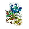
| ||||||||||||
|---|---|---|---|---|---|---|---|---|---|---|---|---|---|
| 1 |
| ||||||||||||
| Unit cell |
|
-Components
| #1: Protein | Mass: 42879.867 Da / Num. of mol.: 1 Source method: isolated from a genetically manipulated source Source: (gene. exp.) Homo sapiens (human) / Gene: CSNK2A2, CK2A2 / Production host:  References: UniProt: P19784, non-specific serine/threonine protein kinase | ||||||||
|---|---|---|---|---|---|---|---|---|---|
| #2: Chemical | ChemComp-EDO /   Mass: 62.068 Da / Num. of mol.: 12 / Source method: obtained synthetically / Formula: C2H6O2 Mass: 62.068 Da / Num. of mol.: 12 / Source method: obtained synthetically / Formula: C2H6O2#3: Chemical | ChemComp-42J / | 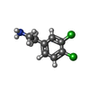  Mass: 190.070 Da / Num. of mol.: 1 / Source method: obtained synthetically / Formula: C8H9Cl2N / Feature type: SUBJECT OF INVESTIGATION Mass: 190.070 Da / Num. of mol.: 1 / Source method: obtained synthetically / Formula: C8H9Cl2N / Feature type: SUBJECT OF INVESTIGATION#4: Chemical | ChemComp-3NG / | 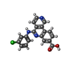  Mass: 349.770 Da / Num. of mol.: 1 / Source method: obtained synthetically / Formula: C19H12ClN3O2 / Feature type: SUBJECT OF INVESTIGATION Mass: 349.770 Da / Num. of mol.: 1 / Source method: obtained synthetically / Formula: C19H12ClN3O2 / Feature type: SUBJECT OF INVESTIGATION#5: Water | ChemComp-HOH / |  Mass: 18.015 Da / Num. of mol.: 282 / Source method: isolated from a natural source / Formula: H2O Mass: 18.015 Da / Num. of mol.: 282 / Source method: isolated from a natural source / Formula: H2OHas ligand of interest | Y | |
-Experimental details
-Experiment
| Experiment | Method: X-RAY DIFFRACTION / Number of used crystals: 1 |
|---|
- Sample preparation
| Crystal | Density Matthews: 2.6 Å3/Da / Density % sol: 52.69 % |
|---|---|
| Crystal grow | Temperature: 293 K / Method: vapor diffusion, sitting drop / pH: 8.5 Details: THE CK2ALPHA' SOLUTION AFTER PROTEIN PURIFICATION (5 MG/ML CK2ALPHA' IN 500 MM NACL, 25 MM TRIS/HCl, PH 8.5) WAS MIXED IN 1:10 VOLUME RATIO WITH 10 MM SOLUTION OF THE INHIBITOR MB002 IN DMSO. ...Details: THE CK2ALPHA' SOLUTION AFTER PROTEIN PURIFICATION (5 MG/ML CK2ALPHA' IN 500 MM NACL, 25 MM TRIS/HCl, PH 8.5) WAS MIXED IN 1:10 VOLUME RATIO WITH 10 MM SOLUTION OF THE INHIBITOR MB002 IN DMSO. THIS SOLUTION WAS MIXED IN 2:1 RATIO WITH RESERVOIR SOLUTION [900 MM LICL, 28 % (W/V) PEG 6000, 250 MM TRIS/HCL, PH 8.5] IN SITTING DROP PLATES (VAPOUR DIFFUSION). THE CRYSTAL GROWTH WAS INDUCED BY MICROSEEDING. THE CRYSTALS WERE OPTIMIZED BY MACROSEEDING. A CRYSTAL WAS PURGED TWICE WITH RESERVOIR SOLUTION BEFORE ADDING 1 MIKROLITER DPA (50 MM IN DMSO) AND 1 MIKROLITER CX-4945 (10 MM IN DMSO) TO THE CRYSTAL MOTHOR LIQUOR FOR EXTENSIVE SOAKING AND REPLACEMENT OF THE INITIAL INHIBITOR MB002. ALL STEPS WERE PERFORMED AT A TEMPERATURE OF 293 K. |
-Data collection
| Diffraction | Mean temperature: 100 K / Serial crystal experiment: N |
|---|---|
| Diffraction source | Source: SYNCHROTRON / Site: PETRA III, EMBL c/o DESY / Beamline: P13 (MX1) / Wavelength: 0.8566 Å |
| Detector | Type: DECTRIS EIGER X 16M / Detector: PIXEL / Date: Dec 16, 2021 |
| Radiation | Protocol: SINGLE WAVELENGTH / Monochromatic (M) / Laue (L): M / Scattering type: x-ray |
| Radiation wavelength | Wavelength: 0.8566 Å / Relative weight: 1 |
| Reflection | Resolution: 1.09→46.17 Å / Num. obs: 118499 / % possible obs: 72.9 % / Redundancy: 6.5 % / Biso Wilson estimate: 11.97 Å2 / CC1/2: 0.997 / Net I/σ(I): 6.5 |
| Reflection shell | Resolution: 1.095→1.134 Å / Num. unique obs: 1763 / CC1/2: 0.608 |
- Processing
| Software |
| |||||||||||||||||||||||||||||||||||||||||||||||||||||||||||||||||||||||||||||||||||||||||||||||||||||||||
|---|---|---|---|---|---|---|---|---|---|---|---|---|---|---|---|---|---|---|---|---|---|---|---|---|---|---|---|---|---|---|---|---|---|---|---|---|---|---|---|---|---|---|---|---|---|---|---|---|---|---|---|---|---|---|---|---|---|---|---|---|---|---|---|---|---|---|---|---|---|---|---|---|---|---|---|---|---|---|---|---|---|---|---|---|---|---|---|---|---|---|---|---|---|---|---|---|---|---|---|---|---|---|---|---|---|---|
| Refinement | Method to determine structure: MOLECULAR REPLACEMENT / Resolution: 1.09→46.17 Å / SU ML: 0.0899 / Cross valid method: FREE R-VALUE / σ(F): 1.96 / Phase error: 23.7104 Stereochemistry target values: GeoStd + Monomer Library + CDL v1.2
| |||||||||||||||||||||||||||||||||||||||||||||||||||||||||||||||||||||||||||||||||||||||||||||||||||||||||
| Solvent computation | Shrinkage radii: 0.9 Å / VDW probe radii: 1.1 Å / Solvent model: FLAT BULK SOLVENT MODEL | |||||||||||||||||||||||||||||||||||||||||||||||||||||||||||||||||||||||||||||||||||||||||||||||||||||||||
| Displacement parameters | Biso mean: 18.8 Å2 | |||||||||||||||||||||||||||||||||||||||||||||||||||||||||||||||||||||||||||||||||||||||||||||||||||||||||
| Refinement step | Cycle: LAST / Resolution: 1.09→46.17 Å
| |||||||||||||||||||||||||||||||||||||||||||||||||||||||||||||||||||||||||||||||||||||||||||||||||||||||||
| Refine LS restraints |
| |||||||||||||||||||||||||||||||||||||||||||||||||||||||||||||||||||||||||||||||||||||||||||||||||||||||||
| LS refinement shell |
|
