 Movie
Movie Controller

[English] 日本語
 Yorodumi
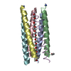
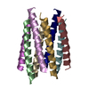
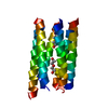
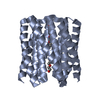
- PDB-8qaa: X-ray crystal structure of a de novo designed antiparallel coiled... -
+ Open data

- Basic information
| Entry | Database: PDB / ID: 8qaa | ||||||
|---|---|---|---|---|---|---|---|
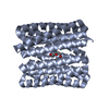
| Title | X-ray crystal structure of a de novo designed antiparallel coiled-coil 6-helix bundle with 4 heptad repeats, antiparallel 6-helix bundle-ALIA | ||||||
 Components Components | antiparallel 6-helix bundle-ALIA | ||||||
 Keywords Keywords | DE NOVO PROTEIN / COILED COIL / 6-HELIX BUNDLE / DE NOVO PROTEIN DESIGN / PEPTIDE ASSEMBLY | ||||||
| Biological species | synthetic construct (others) | ||||||
| Method |  X-RAY DIFFRACTION / SYNCHROTRON / AB INITIO PHASING / Resolution: 1.6 Å X-RAY DIFFRACTION / SYNCHROTRON / AB INITIO PHASING / Resolution: 1.6 Å | ||||||
 Authors Authors | Albanese, K.I. / Petrenas, R. / Woolfson, D.N. | ||||||
| Funding support |  United Kingdom, 1items United Kingdom, 1items
| ||||||
 Citation Citation | Journal: Nat.Chem.Biol. / Year: 2024 Title: Rationally seeded computational protein design of ɑ-helical barrels. Authors: Albanese, K.I. / Petrenas, R. / Pirro, F. / Naudin, E.A. / Borucu, U. / Dawson, W.M. / Scott, D.A. / Leggett, G.J. / Weiner, O.D. / Oliver, T.A.A. / Woolfson, D.N. | ||||||
| History |
|
- Structure visualization
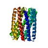
| Structure viewer | Molecule:  MolmilJmol/JSmol MolmilJmol/JSmol |
|---|
- Downloads & links
-Download
| PDBx/mmCIF format | 8qaa.cif.gz | 105 KB | Display | PDBx/mmCIF format |
|---|---|---|---|---|
| PDB format | pdb8qaa.ent.gz | 84 KB | Display | PDB format |
| PDBx/mmJSON format | 8qaa.json.gz | Tree view | PDBx/mmJSON format | |
| Others |  Other downloads Other downloads |
-Validation report
| Summary document | 8qaa_validation.pdf.gz | 423.1 KB | Display | wwPDB validaton report |
|---|---|---|---|---|
| Full document | 8qaa_full_validation.pdf.gz | 423.4 KB | Display | |
| Data in XML | 8qaa_validation.xml.gz | 10.1 KB | Display | |
| Data in CIF | 8qaa_validation.cif.gz | 14.1 KB | Display | |
| Arichive directory | https://data.pdbj.org/pub/pdb/validation_reports/qa/8qaaftp://data.pdbj.org/pub/pdb/validation_reports/qa/8qaa | HTTPS FTP |
-Related structure data


-Links
 PDBj
PDBj
- Assembly
| Deposited unit | 
| ||||||||
|---|---|---|---|---|---|---|---|---|---|
| 1 |
| ||||||||
| Unit cell |
|
-Components
| #1: Protein/peptide | Mass: 3234.871 Da / Num. of mol.: 6 / Source method: obtained synthetically / Source: (synth.) synthetic construct (others) #2: Water | ChemComp-HOH / |  Mass: 18.015 Da / Num. of mol.: 171 / Source method: isolated from a natural source / Formula: H2O Mass: 18.015 Da / Num. of mol.: 171 / Source method: isolated from a natural source / Formula: H2OHas ligand of interest | N | Has protein modification | Y | |
|---|
-Experimental details
-Experiment
| Experiment | Method: X-RAY DIFFRACTION / Number of used crystals: 1 |
|---|
- Sample preparation
| Crystal | Density Matthews: 2 Å3/Da / Density % sol: 38.5 % |
|---|---|
| Crystal grow | Temperature: 293 K / Method: vapor diffusion, sitting drop / pH: 8.5 Details: 12.5% w/v PEG 1000, 12.5% w/v PEG 3350, 12.5% v/v MPD, 0.03 M of each NPS, 0.1 M bicine/Trizma base pH 8.5 |
-Data collection
| Diffraction | Mean temperature: 100 K / Serial crystal experiment: N |
|---|---|
| Diffraction source | Source: SYNCHROTRON / Site: Diamond / Beamline: I04 / Wavelength: 0.98 Å |
| Detector | Type: DECTRIS EIGER2 XE CdTe 16M / Detector: PIXEL / Date: Dec 16, 2021 |
| Radiation | Protocol: SINGLE WAVELENGTH / Monochromatic (M) / Laue (L): M / Scattering type: x-ray |
| Radiation wavelength | Wavelength: 0.98 Å / Relative weight: 1 |
| Reflection | Resolution: 1.6→44.75 Å / Num. obs: 19749 / % possible obs: 98.26 % / Redundancy: 7 % / Biso Wilson estimate: 17.31 Å2 / CC1/2: 0.997 / CC star: 0.999 / Rmerge(I) obs: 0.1184 / Rpim(I) all: 0.0493 / Rrim(I) all: 0.1285 / Net I/σ(I): 8.63 |
| Reflection shell | Resolution: 1.6→1.657 Å / Redundancy: 6.6 % / Rmerge(I) obs: 0.8176 / Mean I/σ(I) obs: 1.11 / Num. unique obs: 19709 / CC1/2: 0.783 / CC star: 0.937 / Rpim(I) all: 0.3437 / Rrim(I) all: 0.8881 / % possible all: 96.93 |
- Processing
| Software |
| |||||||||||||||||||||||||||||||||||||||||||||||||||||||||||||||||||||||||||||||||||||||||||||||||||||||||||||||||||||||||||||||||||||||||||||||||||||||||||||||||||||||||||||||
|---|---|---|---|---|---|---|---|---|---|---|---|---|---|---|---|---|---|---|---|---|---|---|---|---|---|---|---|---|---|---|---|---|---|---|---|---|---|---|---|---|---|---|---|---|---|---|---|---|---|---|---|---|---|---|---|---|---|---|---|---|---|---|---|---|---|---|---|---|---|---|---|---|---|---|---|---|---|---|---|---|---|---|---|---|---|---|---|---|---|---|---|---|---|---|---|---|---|---|---|---|---|---|---|---|---|---|---|---|---|---|---|---|---|---|---|---|---|---|---|---|---|---|---|---|---|---|---|---|---|---|---|---|---|---|---|---|---|---|---|---|---|---|---|---|---|---|---|---|---|---|---|---|---|---|---|---|---|---|---|---|---|---|---|---|---|---|---|---|---|---|---|---|---|---|---|---|
| Refinement | Method to determine structure: AB INITIO PHASING / Resolution: 1.6→44.75 Å / SU ML: 0.19 / Cross valid method: FREE R-VALUE / σ(F): 1.35 / Phase error: 26.08 / Stereochemistry target values: ML
| |||||||||||||||||||||||||||||||||||||||||||||||||||||||||||||||||||||||||||||||||||||||||||||||||||||||||||||||||||||||||||||||||||||||||||||||||||||||||||||||||||||||||||||||
| Solvent computation | Shrinkage radii: 0.9 Å / VDW probe radii: 1.11 Å / Solvent model: FLAT BULK SOLVENT MODEL | |||||||||||||||||||||||||||||||||||||||||||||||||||||||||||||||||||||||||||||||||||||||||||||||||||||||||||||||||||||||||||||||||||||||||||||||||||||||||||||||||||||||||||||||
| Refinement step | Cycle: LAST / Resolution: 1.6→44.75 Å
| |||||||||||||||||||||||||||||||||||||||||||||||||||||||||||||||||||||||||||||||||||||||||||||||||||||||||||||||||||||||||||||||||||||||||||||||||||||||||||||||||||||||||||||||
| Refine LS restraints |
| |||||||||||||||||||||||||||||||||||||||||||||||||||||||||||||||||||||||||||||||||||||||||||||||||||||||||||||||||||||||||||||||||||||||||||||||||||||||||||||||||||||||||||||||
| LS refinement shell |
| |||||||||||||||||||||||||||||||||||||||||||||||||||||||||||||||||||||||||||||||||||||||||||||||||||||||||||||||||||||||||||||||||||||||||||||||||||||||||||||||||||||||||||||||
| Refinement TLS params. | Method: refined / Refine-ID: X-RAY DIFFRACTION
| |||||||||||||||||||||||||||||||||||||||||||||||||||||||||||||||||||||||||||||||||||||||||||||||||||||||||||||||||||||||||||||||||||||||||||||||||||||||||||||||||||||||||||||||
| Refinement TLS group |
|
