 Movie
Movie Controller

[English] 日本語
 Yorodumi
- PDB-8q2o: Structure of alginate transporter AlgE from P. aeruginosa PAO1 by... -
+ Open data

- Basic information
| Entry | Database: PDB / ID: 8q2o | |||||||||
|---|---|---|---|---|---|---|---|---|---|---|
| Title | Structure of alginate transporter AlgE from P. aeruginosa PAO1 by using Se-MAG for the the lipid cubic phase crystallization | |||||||||
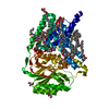
 Components Components | Alginate production protein AlgE | |||||||||
 Keywords Keywords | TRANSPORT PROTEIN / alginate transporter / co-crystallization / experimental phasing / seleno-monoacylglyceride / X-ray scattering | |||||||||
| Function / homology | Alginate export domain / : / Alginate export / alginic acid biosynthetic process / cell outer membrane / CITRATE ANION / : / Alginate production protein AlgE Function and homology information Function and homology information | |||||||||
| Biological species |  Pseudomonas aeruginosa PAO1 (bacteria) Pseudomonas aeruginosa PAO1 (bacteria) | |||||||||
| Method |  X-RAY DIFFRACTION / SYNCHROTRON / SAD / Resolution: 1.7 Å X-RAY DIFFRACTION / SYNCHROTRON / SAD / Resolution: 1.7 Å | |||||||||
 Authors Authors | Huang, C.-Y. / Boland, C. / Kaki, S.S. / Wang, M. / Olieric, V. / Caffrey, M. | |||||||||
| Funding support | European Union,  Ireland, 2items Ireland, 2items
| |||||||||
 Citation Citation | Journal: Crystals / Year: 2023 Title: Se-MAG Is a Convenient Additive for Experimental Phasing and Structure Determination of Membrane Proteins Crystallised by the Lipid Cubic Phase (In Meso) Method Authors: Boland, C. / Huang, C.Y. / Shanker Kaki, S. / Wang, M. / Olieric, V. / Caffrey, M. | |||||||||
| History |
|
- Structure visualization
| Structure viewer | Molecule: MolmilJmol/JSmol |
|---|
- Downloads & links
-Download
| PDBx/mmCIF format | 8q2o.cif.gz | 141.1 KB | Display | PDBx/mmCIF format |
|---|---|---|---|---|
| PDB format | pdb8q2o.ent.gz | 99.6 KB | Display | PDB format |
| PDBx/mmJSON format | 8q2o.json.gz | Tree view | PDBx/mmJSON format | |
| Others |  Other downloads Other downloads |
-Validation report
| Arichive directory | https://data.pdbj.org/pub/pdb/validation_reports/q2/8q2oftp://data.pdbj.org/pub/pdb/validation_reports/q2/8q2o | HTTPS FTP |
|---|
-Related structure data
-Links
 PDBj
PDBj


- Assembly
| Deposited unit | 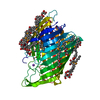
| ||||||||||||
|---|---|---|---|---|---|---|---|---|---|---|---|---|---|
| 1 |
| ||||||||||||
| Unit cell |
|
-Components
-Protein , 1 types, 1 molecules A
| #1: Protein | Mass: 53502.070 Da / Num. of mol.: 1 Source method: isolated from a genetically manipulated source Source: (gene. exp.) Pseudomonas aeruginosa PAO1 (bacteria) / Gene: algE, alg76, PA3544 / Production host: |
|---|
-Non-polymers , 7 types, 341 molecules 

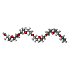


| #2: Chemical | ChemComp-FLC /  Mass: 189.100 Da / Num. of mol.: 1 / Source method: obtained synthetically / Formula: C6H5O7 Mass: 189.100 Da / Num. of mol.: 1 / Source method: obtained synthetically / Formula: C6H5O7 | ||||||||||
|---|---|---|---|---|---|---|---|---|---|---|---|
| #3: Chemical | ChemComp-NA /  Mass: 22.990 Da / Num. of mol.: 5 / Source method: obtained synthetically / Formula: Na Mass: 22.990 Da / Num. of mol.: 5 / Source method: obtained synthetically / Formula: Na#4: Chemical |  Mass: 398.489 Da / Num. of mol.: 2 / Source method: obtained synthetically / Formula: C18H38O9 / Comment: precipitant*YM Mass: 398.489 Da / Num. of mol.: 2 / Source method: obtained synthetically / Formula: C18H38O9 / Comment: precipitant*YM#5: Chemical | ChemComp-IRY / [( Mass: 381.409 Da / Num. of mol.: 16 / Source method: obtained synthetically / Formula: C17H34O4Se / Feature type: SUBJECT OF INVESTIGATION #6: Chemical |  Mass: 306.438 Da / Num. of mol.: 2 / Source method: obtained synthetically / Formula: C16H34O5 / Comment: C8E, detergent*YM Mass: 306.438 Da / Num. of mol.: 2 / Source method: obtained synthetically / Formula: C16H34O5 / Comment: C8E, detergent*YM#7: Chemical | ChemComp-SO4 / |  Mass: 96.063 Da / Num. of mol.: 1 / Source method: obtained synthetically / Formula: SO4 Mass: 96.063 Da / Num. of mol.: 1 / Source method: obtained synthetically / Formula: SO4#8: Water | ChemComp-HOH / | Mass: 18.015 Da / Num. of mol.: 314 / Source method: isolated from a natural source / Formula: H2O |
-Details
| Has ligand of interest | Y |
|---|
-Experimental details
-Experiment
| Experiment | Method: X-RAY DIFFRACTION / Number of used crystals: 1 |
|---|
- Sample preparation
| Crystal | Density Matthews: 2.25 Å3/Da / Density % sol: 45.24 % |
|---|---|
| Crystal grow | Temperature: 293 K / Method: lipidic cubic phase / pH: 5.6 Details: 41 %(v/v) PEG400, 100 mM LiSO4, and 0.1 M sodium citrate at pH 5.6 PH range: 5.6-6.0 |
-Data collection
| Diffraction | Mean temperature: 100 K / Serial crystal experiment: N |
|---|---|
| Diffraction source | Source: SYNCHROTRON / Site: SLS  / Beamline: X06SA / Wavelength: 0.97794 Å / Beamline: X06SA / Wavelength: 0.97794 Å |
| Detector | Type: DECTRIS EIGER X 16M / Detector: PIXEL / Date: Feb 4, 2022 |
| Radiation | Protocol: SINGLE WAVELENGTH / Monochromatic (M) / Laue (L): M / Scattering type: x-ray |
| Radiation wavelength | Wavelength: 0.97794 Å / Relative weight: 1 |
| Reflection | Resolution: 1.7→44.63 Å / Num. obs: 102477 / % possible obs: 100 % / Redundancy: 45.88 % / Biso Wilson estimate: 21.87 Å2 / CC1/2: 0.99 / Rrim(I) all: 0.21 / Net I/σ(I): 17.63 |
| Reflection shell | Resolution: 1.7→1.76 Å / Redundancy: 9.73 % / Mean I/σ(I) obs: 1.61 / Num. unique obs: 7616 / CC1/2: 0.66 / Rrim(I) all: 1.64 / % possible all: 100 |
- Processing
| Software |
| ||||||||||||||||||||||||||||||||||||||||||||||||||||||||||||||||||||||||||||||||||||||||||||||||||||||||||||||||||||||||||||||||||||||||||||
|---|---|---|---|---|---|---|---|---|---|---|---|---|---|---|---|---|---|---|---|---|---|---|---|---|---|---|---|---|---|---|---|---|---|---|---|---|---|---|---|---|---|---|---|---|---|---|---|---|---|---|---|---|---|---|---|---|---|---|---|---|---|---|---|---|---|---|---|---|---|---|---|---|---|---|---|---|---|---|---|---|---|---|---|---|---|---|---|---|---|---|---|---|---|---|---|---|---|---|---|---|---|---|---|---|---|---|---|---|---|---|---|---|---|---|---|---|---|---|---|---|---|---|---|---|---|---|---|---|---|---|---|---|---|---|---|---|---|---|---|---|---|
| Refinement | Method to determine structure: SAD / Resolution: 1.7→44.63 Å / SU ML: 0.2135 / Cross valid method: FREE R-VALUE / σ(F): 1.36 / Phase error: 18.6501 Stereochemistry target values: GeoStd + Monomer Library + CDL v1.2
| ||||||||||||||||||||||||||||||||||||||||||||||||||||||||||||||||||||||||||||||||||||||||||||||||||||||||||||||||||||||||||||||||||||||||||||
| Solvent computation | Shrinkage radii: 0.9 Å / VDW probe radii: 1.1 Å / Solvent model: FLAT BULK SOLVENT MODEL | ||||||||||||||||||||||||||||||||||||||||||||||||||||||||||||||||||||||||||||||||||||||||||||||||||||||||||||||||||||||||||||||||||||||||||||
| Displacement parameters | Biso mean: 27.13 Å2 | ||||||||||||||||||||||||||||||||||||||||||||||||||||||||||||||||||||||||||||||||||||||||||||||||||||||||||||||||||||||||||||||||||||||||||||
| Refinement step | Cycle: LAST / Resolution: 1.7→44.63 Å
| ||||||||||||||||||||||||||||||||||||||||||||||||||||||||||||||||||||||||||||||||||||||||||||||||||||||||||||||||||||||||||||||||||||||||||||
| Refine LS restraints |
| ||||||||||||||||||||||||||||||||||||||||||||||||||||||||||||||||||||||||||||||||||||||||||||||||||||||||||||||||||||||||||||||||||||||||||||
| LS refinement shell |
|
