 Movie
Movie Controller

[English] 日本語
 Yorodumi
- PDB-8q2i: Crystal structure of Ser33 in complex 2HG (2-hydroxyglutarate) an... -
+ Open data

- Basic information
| Entry | Database: PDB / ID: 8q2i | ||||||
|---|---|---|---|---|---|---|---|
| Title | Crystal structure of Ser33 in complex 2HG (2-hydroxyglutarate) and Serine | ||||||
 Components Components | D-3-phosphoglycerate dehydrogenase 2 | ||||||
 Keywords Keywords | CYTOSOLIC PROTEIN / Enzyme protein | ||||||
| Function / homology |  Function and homology information Function and homology information: / 2-oxoglutarate reductase / phosphoglycerate dehydrogenase / phosphoglycerate dehydrogenase activity / L-serine biosynthetic process / NAD binding / identical protein binding / cytoplasm Similarity search - Function | ||||||
| Biological species |  | ||||||
| Method |  X-RAY DIFFRACTION / SYNCHROTRON / MOLECULAR REPLACEMENT / Resolution: 2.51 Å X-RAY DIFFRACTION / SYNCHROTRON / MOLECULAR REPLACEMENT / Resolution: 2.51 Å | ||||||
 Authors Authors | Perrone, S. / Cifuente, J.O. / Marina, A. / Mastrella, L. / Trastoy, B. / Linster, C.L. / Guerin, M.E. | ||||||
| Funding support |  Spain, 1items Spain, 1items
| ||||||
 Citation Citation | Journal: To Be Published Title: Crystal structure of Ser33 in complex 2HG (2-hydroxyglutarate) and Serine Authors: Perrone, S. / Cifuente, J.O. / Marina, A. / Mastrella, L. / Trastoy, B. / Linster, C.L. / Guerin, M.E. | ||||||
| History |
|
- Structure visualization
| Structure viewer | Molecule: MolmilJmol/JSmol |
|---|
- Downloads & links
-Download
| PDBx/mmCIF format | 8q2i.cif.gz | 662.1 KB | Display | PDBx/mmCIF format |
|---|---|---|---|---|
| PDB format | pdb8q2i.ent.gz | 546.7 KB | Display | PDB format |
| PDBx/mmJSON format | 8q2i.json.gz | Tree view | PDBx/mmJSON format | |
| Others |  Other downloads Other downloads |
-Validation report
| Summary document | 8q2i_validation.pdf.gz | 5.7 MB | Display | wwPDB validaton report |
|---|---|---|---|---|
| Full document | 8q2i_full_validation.pdf.gz | 5.7 MB | Display | |
| Data in XML | 8q2i_validation.xml.gz | 138.9 KB | Display | |
| Data in CIF | 8q2i_validation.cif.gz | 175.5 KB | Display | |
| Arichive directory | https://data.pdbj.org/pub/pdb/validation_reports/q2/8q2iftp://data.pdbj.org/pub/pdb/validation_reports/q2/8q2i | HTTPS FTP |
-Related structure data
| Similar structure data |
|---|
-Links
 PDBj
PDBj

- Assembly
| Deposited unit | 
| ||||||||||
|---|---|---|---|---|---|---|---|---|---|---|---|
| 1 | 
| ||||||||||
| 2 | 
| ||||||||||
| Unit cell |
|
-Components
| #1: Protein | Mass: 51243.039 Da / Num. of mol.: 8 Source method: isolated from a genetically manipulated source Source: (gene. exp.) Gene: SER33, YIL074C Production host:  References: UniProt: P40510, phosphoglycerate dehydrogenase, 2-oxoglutarate reductase #2: Chemical | ChemComp-NAD /   Mass: 663.425 Da / Num. of mol.: 8 / Source method: obtained synthetically / Formula: C21H27N7O14P2 / Feature type: SUBJECT OF INVESTIGATION / Comment: NAD*YM Mass: 663.425 Da / Num. of mol.: 8 / Source method: obtained synthetically / Formula: C21H27N7O14P2 / Feature type: SUBJECT OF INVESTIGATION / Comment: NAD*YM#3: Chemical | ChemComp-SER /   Type: L-peptide linking / Mass: 105.093 Da / Num. of mol.: 8 / Source method: obtained synthetically / Formula: C3H7NO3 / Feature type: SUBJECT OF INVESTIGATION Type: L-peptide linking / Mass: 105.093 Da / Num. of mol.: 8 / Source method: obtained synthetically / Formula: C3H7NO3 / Feature type: SUBJECT OF INVESTIGATION#4: Chemical | ChemComp-2HG / ( 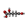  Mass: 148.114 Da / Num. of mol.: 4 / Source method: obtained synthetically / Formula: C5H8O5 / Feature type: SUBJECT OF INVESTIGATION Mass: 148.114 Da / Num. of mol.: 4 / Source method: obtained synthetically / Formula: C5H8O5 / Feature type: SUBJECT OF INVESTIGATION#5: Water | ChemComp-HOH / |  Mass: 18.015 Da / Num. of mol.: 182 / Source method: isolated from a natural source / Formula: H2O Mass: 18.015 Da / Num. of mol.: 182 / Source method: isolated from a natural source / Formula: H2OHas ligand of interest | Y | Has protein modification | N | |
|---|
-Experimental details
-Experiment
| Experiment | Method: X-RAY DIFFRACTION / Number of used crystals: 1 |
|---|
- Sample preparation
| Crystal | Density Matthews: 2.77 Å3/Da / Density % sol: 55.59 % |
|---|---|
| Crystal grow | Temperature: 291 K / Method: vapor diffusion, sitting drop Details: 0.2 M Sodium citrate tribasic dihydrate 0.1 M Bis-Tris propane 8.5 20 % w/v PEG 3350 7.4 mg/ml of protein in 25 mM Tris pH=7.5 and 150 mM NaCl |
-Data collection
| Diffraction | Mean temperature: 100 K / Serial crystal experiment: N |
|---|---|
| Diffraction source | Source: SYNCHROTRON / Site: ALBA / Beamline: XALOC / Wavelength: 0.9792 Å |
| Detector | Type: DECTRIS PILATUS 6M / Detector: PIXEL / Date: Nov 28, 2020 |
| Radiation | Protocol: SINGLE WAVELENGTH / Monochromatic (M) / Laue (L): M / Scattering type: x-ray |
| Radiation wavelength | Wavelength: 0.9792 Å / Relative weight: 1 |
| Reflection | Resolution: 2.51→20.02 Å / Num. obs: 146160 / % possible obs: 97.21 % / Redundancy: 3.5 % / CC1/2: 0.997 / Net I/σ(I): 10.55 |
| Reflection shell | Resolution: 2.51→2.6 Å / Num. unique obs: 14197 / CC1/2: 0.661 |
- Processing
| Software |
| ||||||||||||||||
|---|---|---|---|---|---|---|---|---|---|---|---|---|---|---|---|---|---|
| Refinement | Method to determine structure: MOLECULAR REPLACEMENT / Resolution: 2.51→20.02 Å / Cross valid method: FREE R-VALUE
| ||||||||||||||||
| Refinement step | Cycle: LAST / Resolution: 2.51→20.02 Å
|
