 Movie
Movie Controller

[English] 日本語
 Yorodumi
- PDB-8q20: Crystal structure of Vanadium-dependent haloperoxidase R425D muta... -
+ Open data

- Basic information
| Entry | Database: PDB / ID: 8q20 | ||||||
|---|---|---|---|---|---|---|---|
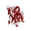
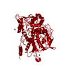
| Title | Crystal structure of Vanadium-dependent haloperoxidase R425D mutant (A. marina) | ||||||
 Components Components | Vanadium-dependent bromoperoxidase, putative | ||||||
 Keywords Keywords | OXIDOREDUCTASE / Vanadium / Enzyme Catalysis / Peroxidase / Halogenation / Mutant | ||||||
| Function / homology | Bromoperoxidase/chloroperoxidase C-terminal / : / Phosphatidic acid phosphatase type 2/haloperoxidase superfamily / peroxidase activity / PHOSPHATE ION / Vanadium-dependent bromoperoxidase, putative Function and homology information Function and homology information | ||||||
| Biological species |  Acaryochloris marina (bacteria) Acaryochloris marina (bacteria) | ||||||
| Method |  X-RAY DIFFRACTION / SYNCHROTRON / MOLECULAR REPLACEMENT / Resolution: 3.5 Å X-RAY DIFFRACTION / SYNCHROTRON / MOLECULAR REPLACEMENT / Resolution: 3.5 Å | ||||||
 Authors Authors | Zeides, P. / Bellmannn-Sickert, K. / Zhang, R. / Seel, C.J. / Most, V. / Schroeder, C.T. / Groll, M. / Gulder, T. | ||||||
| Funding support | 1items
| ||||||
 Citation Citation | Journal: Nat Commun / Year: 2025 Title: Unraveling the molecular basis of substrate specificity and halogen activation in vanadium-dependent haloperoxidases. Authors: Zeides, P. / Bellmann-Sickert, K. / Zhang, R. / Seel, C.J. / Most, V. / Schoeder, C.T. / Groll, M. / Gulder, T. | ||||||
| History |
|
- Structure visualization
| Structure viewer | Molecule: MolmilJmol/JSmol |
|---|
- Downloads & links
-Download
| PDBx/mmCIF format | 8q20.cif.gz | 254.9 KB | Display | PDBx/mmCIF format |
|---|---|---|---|---|
| PDB format | pdb8q20.ent.gz | 207.8 KB | Display | PDB format |
| PDBx/mmJSON format | 8q20.json.gz | Tree view | PDBx/mmJSON format | |
| Others |  Other downloads Other downloads |
-Validation report
| Arichive directory | https://data.pdbj.org/pub/pdb/validation_reports/q2/8q20ftp://data.pdbj.org/pub/pdb/validation_reports/q2/8q20 | HTTPS FTP |
|---|
-Related structure data

-Links
 PDBj
PDBj- Assembly
| Deposited unit | 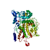
| ||||||||
|---|---|---|---|---|---|---|---|---|---|
| 1 | x 12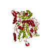
| ||||||||
| Unit cell |
|
-Components
| #1: Protein | Mass: 73068.539 Da / Num. of mol.: 1 / Mutation: R452D Source method: isolated from a genetically manipulated source Source: (gene. exp.) Acaryochloris marina (bacteria) / Gene: AM1_4975 / Plasmid: pET28b / Production host: |
|---|---|
| #2: Chemical | ChemComp-PO4 /   Mass: 94.971 Da / Num. of mol.: 1 / Source method: obtained synthetically / Formula: PO4 / Feature type: SUBJECT OF INVESTIGATION Mass: 94.971 Da / Num. of mol.: 1 / Source method: obtained synthetically / Formula: PO4 / Feature type: SUBJECT OF INVESTIGATION |
| #3: Chemical | ChemComp-SO4 /   Mass: 96.063 Da / Num. of mol.: 1 / Source method: obtained synthetically / Formula: SO4 Mass: 96.063 Da / Num. of mol.: 1 / Source method: obtained synthetically / Formula: SO4 |
| Has ligand of interest | Y |
| Has protein modification | Y |
-Experimental details
-Experiment
| Experiment | Method: X-RAY DIFFRACTION / Number of used crystals: 1 |
|---|
- Sample preparation
| Crystal | Density Matthews: 4.18 Å3/Da / Density % sol: 70.58 % |
|---|---|
| Crystal grow | Temperature: 293 K / Method: vapor diffusion, hanging drop / pH: 8.5 Details: 0.1 M TRIS, 1.25 M Ammoniumdihydrogenphosphate, 0.5 mM potassium vanadate |
-Data collection
| Diffraction | Mean temperature: 100 K / Serial crystal experiment: N |
|---|---|
| Diffraction source | Source: SYNCHROTRON / Site: SLS  / Beamline: X06SA / Wavelength: 1 Å / Beamline: X06SA / Wavelength: 1 Å |
| Detector | Type: DECTRIS EIGER X 16M / Detector: PIXEL / Date: Nov 17, 2019 |
| Radiation | Protocol: SINGLE WAVELENGTH / Monochromatic (M) / Laue (L): M / Scattering type: x-ray |
| Radiation wavelength | Wavelength: 1 Å / Relative weight: 1 |
| Reflection | Resolution: 3.5→30 Å / Num. obs: 15830 / % possible obs: 96.5 % / Redundancy: 5 % / Rmerge(I) obs: 0.122 / Net I/σ(I): 9.4 |
| Reflection shell | Resolution: 3.5→3.6 Å / Redundancy: 5 % / Rmerge(I) obs: 0.632 / Num. unique obs: 1244 / % possible all: 96.6 |
- Processing
| Software |
| |||||||||||||||||||||||||||||||||||||||||||||||||||||||||||||||||
|---|---|---|---|---|---|---|---|---|---|---|---|---|---|---|---|---|---|---|---|---|---|---|---|---|---|---|---|---|---|---|---|---|---|---|---|---|---|---|---|---|---|---|---|---|---|---|---|---|---|---|---|---|---|---|---|---|---|---|---|---|---|---|---|---|---|---|
| Refinement | Method to determine structure: MOLECULAR REPLACEMENT / Resolution: 3.5→30 Å / Cor.coef. Fo:Fc: 0.931 / Cor.coef. Fo:Fc free: 0.926 / SU B: 54.558 / SU ML: 0.361 / Cross valid method: THROUGHOUT / σ(F): 0 / ESU R Free: 0.502 / Stereochemistry target values: MAXIMUM LIKELIHOOD Details: HYDROGENS HAVE BEEN ADDED IN THE RIDING POSITIONS U VALUES : WITH TLS ADDED
| |||||||||||||||||||||||||||||||||||||||||||||||||||||||||||||||||
| Solvent computation | Ion probe radii: 0.8 Å / Shrinkage radii: 0.8 Å / VDW probe radii: 1.2 Å / Solvent model: MASK | |||||||||||||||||||||||||||||||||||||||||||||||||||||||||||||||||
| Displacement parameters | Biso max: 160.04 Å2 / Biso mean: 96.498 Å2 / Biso min: 81.59 Å2
| |||||||||||||||||||||||||||||||||||||||||||||||||||||||||||||||||
| Refinement step | Cycle: final / Resolution: 3.5→30 Å
| |||||||||||||||||||||||||||||||||||||||||||||||||||||||||||||||||
| Refine LS restraints |
| |||||||||||||||||||||||||||||||||||||||||||||||||||||||||||||||||
| LS refinement shell | Resolution: 3.5→3.59 Å / Rfactor Rfree error: 0 / Total num. of bins used: 20
| |||||||||||||||||||||||||||||||||||||||||||||||||||||||||||||||||
| Refinement TLS params. | Method: refined / Origin x: 19.9442 Å / Origin y: 4.7943 Å / Origin z: 44.8523 Å
|
