 Movie
Movie Controller

[English] 日本語
 Yorodumi
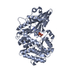
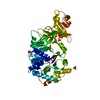
- PDB-8pyx: Amide bond synthetase from Streptomyces hindustanus K492H mutant ... -
+ Open data

- Basic information
| Entry | Database: PDB / ID: 8pyx | ||||||
|---|---|---|---|---|---|---|---|
| Title | Amide bond synthetase from Streptomyces hindustanus K492H mutant in complex with Adenosine | ||||||
 Components Components | Fatty-acyl-CoA synthase | ||||||
 Keywords Keywords | LIGASE / Amide / Amide Bond Synthetase / ATP | ||||||
| Function / homology |  Function and homology information Function and homology information | ||||||
| Biological species |  Streptoalloteichus hindustanus (bacteria) Streptoalloteichus hindustanus (bacteria) | ||||||
| Method |  X-RAY DIFFRACTION / SYNCHROTRON / MOLECULAR REPLACEMENT / Resolution: 2.02 Å X-RAY DIFFRACTION / SYNCHROTRON / MOLECULAR REPLACEMENT / Resolution: 2.02 Å | ||||||
 Authors Authors | Tang, Q. / Grogan, G. | ||||||
| Funding support |  United Kingdom, 1items United Kingdom, 1items
| ||||||
 Citation Citation | Journal: Acs Catalysis / Year: 2024 Title: Broad Spectrum Enantioselective Amide Bond Synthetase from Streptoalloteichus hindustanus. Authors: Tang, Q. / Petchey, M. / Rowlinson, B. / Burden, T.J. / Fairlamb, I.J.S. / Grogan, G. | ||||||
| History |
|
- Structure visualization
| Structure viewer | Molecule: MolmilJmol/JSmol |
|---|
- Downloads & links
-Download
| PDBx/mmCIF format | 8pyx.cif.gz | 208.7 KB | Display | PDBx/mmCIF format |
|---|---|---|---|---|
| PDB format | pdb8pyx.ent.gz | 162.5 KB | Display | PDB format |
| PDBx/mmJSON format | 8pyx.json.gz | Tree view | PDBx/mmJSON format | |
| Others |  Other downloads Other downloads |
-Validation report
| Summary document | 8pyx_validation.pdf.gz | 1.1 MB | Display | wwPDB validaton report |
|---|---|---|---|---|
| Full document | 8pyx_full_validation.pdf.gz | 1.1 MB | Display | |
| Data in XML | 8pyx_validation.xml.gz | 40.8 KB | Display | |
| Data in CIF | 8pyx_validation.cif.gz | 59.3 KB | Display | |
| Arichive directory | https://data.pdbj.org/pub/pdb/validation_reports/py/8pyxftp://data.pdbj.org/pub/pdb/validation_reports/py/8pyx | HTTPS FTP |
-Related structure data
-Links
 PDBj
PDBj

- Assembly
| Deposited unit | 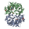
| ||||||||
|---|---|---|---|---|---|---|---|---|---|
| 1 | 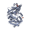
| ||||||||
| 2 | 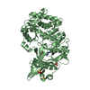
| ||||||||
| Unit cell |
|
-Components
| #1: Protein | Mass: 54450.410 Da / Num. of mol.: 2 / Mutation: K492H Source method: isolated from a genetically manipulated source Source: (gene. exp.) Streptoalloteichus hindustanus (bacteria)Gene: SAMN05444320_10350 / Production host: #2: Chemical | ChemComp-SO4 /   Mass: 96.063 Da / Num. of mol.: 6 / Source method: obtained synthetically / Formula: SO4 Mass: 96.063 Da / Num. of mol.: 6 / Source method: obtained synthetically / Formula: SO4#3: Chemical |   Mass: 267.241 Da / Num. of mol.: 2 / Source method: obtained synthetically / Formula: C10H13N5O4 / Feature type: SUBJECT OF INVESTIGATION Mass: 267.241 Da / Num. of mol.: 2 / Source method: obtained synthetically / Formula: C10H13N5O4 / Feature type: SUBJECT OF INVESTIGATION#4: Water | ChemComp-HOH / |  Mass: 18.015 Da / Num. of mol.: 506 / Source method: isolated from a natural source / Formula: H2O Mass: 18.015 Da / Num. of mol.: 506 / Source method: isolated from a natural source / Formula: H2OHas ligand of interest | Y | |
|---|
-Experimental details
-Experiment
| Experiment | Method: X-RAY DIFFRACTION / Number of used crystals: 1 |
|---|
- Sample preparation
| Crystal | Density Matthews: 2.8 Å3/Da / Density % sol: 56.13 % |
|---|---|
| Crystal grow | Temperature: 277 K / Method: vapor diffusion, sitting drop / pH: 7.5 / Details: 0.2 M Li2SO4; 0.1 M HEPES pH 7.5; 25% PEG 3350 |
-Data collection
| Diffraction | Mean temperature: 120 K / Serial crystal experiment: N |
|---|---|
| Diffraction source | Source: SYNCHROTRON / Site: Diamond / Beamline: I03 / Wavelength: 0.95881 Å |
| Detector | Type: DECTRIS EIGER2 XE 16M / Detector: PIXEL / Date: Sep 16, 2022 |
| Radiation | Protocol: SINGLE WAVELENGTH / Monochromatic (M) / Laue (L): M / Scattering type: x-ray |
| Radiation wavelength | Wavelength: 0.95881 Å / Relative weight: 1 |
| Reflection | Resolution: 2.02→29.35 Å / Num. obs: 78528 / % possible obs: 99 % / Redundancy: 7 % / Biso Wilson estimate: 26 Å2 / CC1/2: 1 / Rmerge(I) obs: 0.12 / Rpim(I) all: 0.06 / Net I/σ(I): 11.6 |
| Reflection shell | Resolution: 2.02→2.06 Å / Rmerge(I) obs: 1.05 / Mean I/σ(I) obs: 1.7 / Num. unique obs: 3766 / CC1/2: 0.8 / Rpim(I) all: 0.55 |
- Processing
| Software |
| ||||||||||||||||||||||||||||||||||||||||||||||||||||||||||||||||||||||||||||||||||||||||||||||||||||||||||||||||||||||||||||||||||||||||||||||||||||||||||||||||||||||||||||||||||||||
|---|---|---|---|---|---|---|---|---|---|---|---|---|---|---|---|---|---|---|---|---|---|---|---|---|---|---|---|---|---|---|---|---|---|---|---|---|---|---|---|---|---|---|---|---|---|---|---|---|---|---|---|---|---|---|---|---|---|---|---|---|---|---|---|---|---|---|---|---|---|---|---|---|---|---|---|---|---|---|---|---|---|---|---|---|---|---|---|---|---|---|---|---|---|---|---|---|---|---|---|---|---|---|---|---|---|---|---|---|---|---|---|---|---|---|---|---|---|---|---|---|---|---|---|---|---|---|---|---|---|---|---|---|---|---|---|---|---|---|---|---|---|---|---|---|---|---|---|---|---|---|---|---|---|---|---|---|---|---|---|---|---|---|---|---|---|---|---|---|---|---|---|---|---|---|---|---|---|---|---|---|---|---|---|
| Refinement | Method to determine structure: MOLECULAR REPLACEMENT / Resolution: 2.02→29.35 Å / Cor.coef. Fo:Fc: 0.956 / Cor.coef. Fo:Fc free: 0.94 / SU B: 5.316 / SU ML: 0.143 / Cross valid method: THROUGHOUT / ESU R: 0.2 / ESU R Free: 0.177 / Stereochemistry target values: MAXIMUM LIKELIHOOD / Details: HYDROGENS HAVE BEEN ADDED IN THE RIDING POSITIONS
| ||||||||||||||||||||||||||||||||||||||||||||||||||||||||||||||||||||||||||||||||||||||||||||||||||||||||||||||||||||||||||||||||||||||||||||||||||||||||||||||||||||||||||||||||||||||
| Solvent computation | Ion probe radii: 0.8 Å / Shrinkage radii: 0.8 Å / VDW probe radii: 1.2 Å / Solvent model: MASK | ||||||||||||||||||||||||||||||||||||||||||||||||||||||||||||||||||||||||||||||||||||||||||||||||||||||||||||||||||||||||||||||||||||||||||||||||||||||||||||||||||||||||||||||||||||||
| Displacement parameters | Biso mean: 30.238 Å2
| ||||||||||||||||||||||||||||||||||||||||||||||||||||||||||||||||||||||||||||||||||||||||||||||||||||||||||||||||||||||||||||||||||||||||||||||||||||||||||||||||||||||||||||||||||||||
| Refinement step | Cycle: 1 / Resolution: 2.02→29.35 Å
| ||||||||||||||||||||||||||||||||||||||||||||||||||||||||||||||||||||||||||||||||||||||||||||||||||||||||||||||||||||||||||||||||||||||||||||||||||||||||||||||||||||||||||||||||||||||
| Refine LS restraints |
|