Protein / Protein/peptide / Sugars , 3 types, 3 molecules AB
#1: Protein
L,D-transpeptidase2 / LDT 2 / Ldt(Mt2)
Mass: 37993.082 Da / Num. of mol.: 1 Source method: isolated from a genetically manipulated source Source: (gene. exp.) Mycobacterium tuberculosis (bacteria) / Gene: ldtB, MT2594, V735_02606 / Production host: Escherichia coli BL21(DE3) (bacteria) References: UniProt: O53223, Transferases; Acyltransferases; Aminoacyltransferases
#2: Protein/peptide
Peptidoglycantetrapeptide
Type: Glycopeptide / Class: Inducer / Mass: 459.496 Da / Num. of mol.: 1 / Source method: isolated from a natural source Details: Peptidoglycan is composed of glycan strands and peptide stems. The glycan strands are made of N-acetylmuramic acid/MurNAc (MUB) and N-acetylglucosamine/GlcNAc (NAG) linked by beta-1,4 ...Details: Peptidoglycan is composed of glycan strands and peptide stems. The glycan strands are made of N-acetylmuramic acid/MurNAc (MUB) and N-acetylglucosamine/GlcNAc (NAG) linked by beta-1,4 glycosidic bonds. Peptide stems, which are less conserved, attach to MurNAc and consist of L-alanine, gamma D-glutamine/D-Gln (ZGL), meso-diaminopimelic acid/m-DAP (JGO), and D-alanine (DAP). Source: (natural) Corynebacterium jeikeium (bacteria) / References: BIRD: PRD_002583
Type: oligosaccharide, Glycopeptide / Class: Inducer / Mass: 496.463 Da / Num. of mol.: 1 Source method: isolated from a genetically manipulated source Details: Peptidoglycan is composed of glycan strands and peptide stems. The glycan strands are made of N-acetylmuramic acid/MurNAc (MUB) and N-acetylglucosamine/GlcNAc (NAG) linked by beta-1,4 ...Details: Peptidoglycan is composed of glycan strands and peptide stems. The glycan strands are made of N-acetylmuramic acid/MurNAc (MUB) and N-acetylglucosamine/GlcNAc (NAG) linked by beta-1,4 glycosidic bonds. Peptide stems, which are less conserved, attach to MurNAc and consist of L-alanine, gamma D-glutamine/D-Gln (ZGL), meso-diaminopimelic acid/m-DAP (JGO), and D-alanine (DAP). References: BIRD: PRD_002583
In the structure databanks used in Yorodumi, some data are registered as the other names, "COVID-19 virus" and "2019-nCoV". Here are the details of the virus and the list of structure data.
Jan 31, 2019. EMDB accession codes are about to change! (news from PDBe EMDB page)
EMDB accession codes are about to change! (news from PDBe EMDB page)
The allocation of 4 digits for EMDB accession codes will soon come to an end. Whilst these codes will remain in use, new EMDB accession codes will include an additional digit and will expand incrementally as the available range of codes is exhausted. The current 4-digit format prefixed with “EMD-” (i.e. EMD-XXXX) will advance to a 5-digit format (i.e. EMD-XXXXX), and so on. It is currently estimated that the 4-digit codes will be depleted around Spring 2019, at which point the 5-digit format will come into force.
The EM Navigator/Yorodumi systems omit the EMD- prefix.
Related info.:Q: What is EMD? / ID/Accession-code notation in Yorodumi/EM Navigator
Yorodumi is a browser for structure data from EMDB, PDB, SASBDB, etc.
This page is also the successor to EM Navigator detail page, and also detail information page/front-end page for Omokage search.
The word "yorodu" (or yorozu) is an old Japanese word meaning "ten thousand". "mi" (miru) is to see.
Related info.:EMDB / PDB / SASBDB / Comparison of 3 databanks / Yorodumi Search / Aug 31, 2016. New EM Navigator & Yorodumi / Yorodumi Papers / Jmol/JSmol / Function and homology information / Changes in new EM Navigator and Yorodumi
 Movie
Movie Controller

 Yorodumi
 Open data
 Basic information
 Components
 Keywords
 Function and homology information
 Mycobacterium tuberculosis (bacteria)
 X-RAY DIFFRACTION /
  Authors
 United Kingdom, 1items
  Citation
 Structure visualization
 Downloads & links
 Other downloads
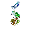
 PDBj
PDBj


 Assembly


 Mass: 238.305 Da / Num. of mol.: 1 / Source method: obtained synthetically / Formula: C8H18N2O4S / Comment: pH buffer*YM
 Mass: 35.453 Da / Num. of mol.: 1 / Source method: obtained synthetically / Formula: Cl
 Sample preparation
 Processing