 Movie
Movie Controller


+ Open data

- Basic information
| Entry | Database: PDB / ID: 8pts | ||||||
|---|---|---|---|---|---|---|---|
| Title | human GUK1 in complex with compound AT8001 | ||||||
 Components Components | Guanylate kinase | ||||||
 Keywords Keywords | CONTRACTILE PROTEIN / complex intermediate compound inhibitor / kinase / Guanylate kinase | ||||||
| Function / homology |  Function and homology information Function and homology informationdGDP biosynthetic process / dATP metabolic process / GDP biosynthetic process / GDP-mannose metabolic process / guanylate kinase / dGMP metabolic process / glycoprotein transport / purine nucleotide metabolic process / GMP kinase activity / nucleobase-containing small molecule interconversion ...dGDP biosynthetic process / dATP metabolic process / GDP biosynthetic process / GDP-mannose metabolic process / guanylate kinase / dGMP metabolic process / glycoprotein transport / purine nucleotide metabolic process / GMP kinase activity / nucleobase-containing small molecule interconversion / Interconversion of nucleotide di- and triphosphates / Azathioprine ADME / photoreceptor inner segment / xenobiotic metabolic process / mitochondrion / ATP binding / cytosol Similarity search - Function | ||||||
| Biological species |  Homo sapiens (human) Homo sapiens (human) | ||||||
| Method |  X-RAY DIFFRACTION / SYNCHROTRON / MOLECULAR REPLACEMENT / Resolution: 1.76 Å X-RAY DIFFRACTION / SYNCHROTRON / MOLECULAR REPLACEMENT / Resolution: 1.76 Å | ||||||
 Authors Authors | Zimberger, C. / Canard, B. / Ferron, F. | ||||||
| Funding support |  France, 1items France, 1items
| ||||||
 Citation Citation | Journal: Plos Biol. / Year: 2024 Title: The activation cascade of the broad-spectrum antiviral bemnifosbuvir characterized at atomic resolution. Authors: Chazot, A. / Zimberger, C. / Feracci, M. / Moussa, A. / Good, S. / Sommadossi, J.P. / Alvarez, K. / Ferron, F. / Canard, B. #1: Journal: Biorxiv / Year: 2024Title: The activation chain of the broad-spectrum antiviral bemnifosbuvir at atomic resolution Authors: Chazot, A. / Zimberger, C. / Feracci, M. / Moussa, A. / Good, S. / Sommadossi, J.P. / Alvarez, K. / Ferron, F. / Canard, B. | ||||||
| History |
|
- Structure visualization
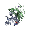
| Structure viewer | Molecule: MolmilJmol/JSmol |
|---|
- Downloads & links
-Download
| PDBx/mmCIF format | 8pts.cif.gz | 164.6 KB | Display | PDBx/mmCIF format |
|---|---|---|---|---|
| PDB format | pdb8pts.ent.gz | 132 KB | Display | PDB format |
| PDBx/mmJSON format | 8pts.json.gz | Tree view | PDBx/mmJSON format | |
| Others |  Other downloads Other downloads |
-Validation report
| Arichive directory | https://data.pdbj.org/pub/pdb/validation_reports/pt/8ptsftp://data.pdbj.org/pub/pdb/validation_reports/pt/8pts | HTTPS FTP |
|---|
-Related structure data
-Links
 PDBj
PDBj

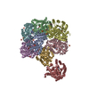
- Assembly
| Deposited unit | 
| ||||||||
|---|---|---|---|---|---|---|---|---|---|
| 1 |
| ||||||||
| Unit cell |
| ||||||||
| Components on special symmetry positions |
|
-Components
| #1: Protein | Mass: 21753.584 Da / Num. of mol.: 2 Source method: isolated from a genetically manipulated source Source: (gene. exp.) Homo sapiens (human) / Gene: GUK1, GMK, GMPKProduction host:  References: UniProt: Q16774, guanylate kinase #2: Chemical | Mass: 379.238 Da / Num. of mol.: 2 / Source method: obtained synthetically / Formula: C11H15FN5O7P / Feature type: SUBJECT OF INVESTIGATION #3: Chemical | ChemComp-GOL / |   Mass: 92.094 Da / Num. of mol.: 1 / Source method: obtained synthetically / Formula: C3H8O3 Mass: 92.094 Da / Num. of mol.: 1 / Source method: obtained synthetically / Formula: C3H8O3#4: Water | ChemComp-HOH / |  Mass: 18.015 Da / Num. of mol.: 119 / Source method: isolated from a natural source / Formula: H2O Mass: 18.015 Da / Num. of mol.: 119 / Source method: isolated from a natural source / Formula: H2OHas ligand of interest | Y | |
|---|
-Experimental details
-Experiment
| Experiment | Method: X-RAY DIFFRACTION / Number of used crystals: 1 |
|---|
- Sample preparation
| Crystal | Density Matthews: 2.31 Å3/Da / Density % sol: 46.78 % |
|---|---|
| Crystal grow | Temperature: 293.15 K / Method: vapor diffusion, sitting drop Details: 12 -17 % PEG330 66mM Na Cacodylate 33mM MES 66mM MgCl2 PH range: 6.5-7.5 |
-Data collection
| Diffraction | Mean temperature: 100 K / Serial crystal experiment: N |
|---|---|
| Diffraction source | Source: SYNCHROTRON / Site: ESRF / Beamline: ID23-1 / Wavelength: 0.8856 Å |
| Detector | Type: DECTRIS PILATUS 6M / Detector: PIXEL / Date: Sep 8, 2022 |
| Radiation | Protocol: SINGLE WAVELENGTH / Monochromatic (M) / Laue (L): M / Scattering type: x-ray |
| Radiation wavelength | Wavelength: 0.8856 Å / Relative weight: 1 |
| Reflection | Resolution: 1.76→62.849 Å / Num. obs: 37848 / % possible obs: 96 % / Redundancy: 4.2 % / CC1/2: 0.998 / Rmerge(I) obs: 0.055 / Rpim(I) all: 0.029 / Rrim(I) all: 0.063 / Net I/σ(I): 9.7 |
| Reflection shell | Resolution: 1.763→1.793 Å / Redundancy: 4.3 % / Rmerge(I) obs: 0.699 / Mean I/σ(I) obs: 2.2 / Num. unique obs: 1925 / CC1/2: 0.825 / Rpim(I) all: 0.371 / Rrim(I) all: 0.796 / % possible all: 96.2 |
- Processing
| Software |
| ||||||||||||||||||||||||||||||||||||||||
|---|---|---|---|---|---|---|---|---|---|---|---|---|---|---|---|---|---|---|---|---|---|---|---|---|---|---|---|---|---|---|---|---|---|---|---|---|---|---|---|---|---|
| Refinement | Method to determine structure: MOLECULAR REPLACEMENT / Resolution: 1.76→62.849 Å / Cross valid method: THROUGHOUT Details: HYDROGENS WERE FULLY REFINED WITH ZERO OCCUPANCY AT NUCLEAR POSITION.
| ||||||||||||||||||||||||||||||||||||||||
| Refinement step | Cycle: LAST / Resolution: 1.76→62.849 Å
| ||||||||||||||||||||||||||||||||||||||||
| Refine LS restraints |
|
