 Movie
Movie Controller


+ Open data

- Basic information
| Entry | Database: PDB / ID: 8prd | ||||||
|---|---|---|---|---|---|---|---|
| Title | The structure of nvBagel2 | ||||||
 Components Components | Cell surface protein | ||||||
 Keywords Keywords | BIOSYNTHETIC PROTEIN / Protein design / symmetric / assembly / self-assembly / beta-propeller | ||||||
| Function / homology |  Function and homology information Function and homology informationamino acid activation for nonribosomal peptide biosynthetic process / metal ion binding Similarity search - Function | ||||||
| Biological species |   Dictyoglomus thermophilum (bacteria) Dictyoglomus thermophilum (bacteria) | ||||||
| Method |  X-RAY DIFFRACTION / SYNCHROTRON / MOLECULAR REPLACEMENT / Resolution: 1.41 Å X-RAY DIFFRACTION / SYNCHROTRON / MOLECULAR REPLACEMENT / Resolution: 1.41 Å | ||||||
 Authors Authors | Vandebroek, L. / Voet, A.R.D. / Lee, X.Y. | ||||||
| Funding support |  Belgium, 1items Belgium, 1items
| ||||||
 Citation Citation | Journal: To Be Published Title: The structure of v13Bagel2 Authors: Vandebroek, L. / Voet, A.R.D. / Lee, X.Y. | ||||||
| History |
|
- Structure visualization
| Structure viewer | Molecule: MolmilJmol/JSmol |
|---|
- Downloads & links
-Download
| PDBx/mmCIF format | 8prd.cif.gz | 57.6 KB | Display | PDBx/mmCIF format |
|---|---|---|---|---|
| PDB format | pdb8prd.ent.gz | 41.5 KB | Display | PDB format |
| PDBx/mmJSON format | 8prd.json.gz | Tree view | PDBx/mmJSON format | |
| Others |  Other downloads Other downloads |
-Validation report
| Arichive directory | https://data.pdbj.org/pub/pdb/validation_reports/pr/8prdftp://data.pdbj.org/pub/pdb/validation_reports/pr/8prd | HTTPS FTP |
|---|
-Related structure data
| Related structure data |  8pr9C 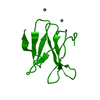 8preC 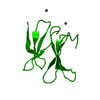 8prfC 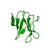 8prgC 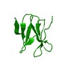 8prhC 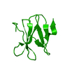 8priC 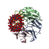 8prmC 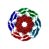 8proC C: citing same article ( |
|---|---|
| Similar structure data |
-Links
 PDBj
PDBj


- Assembly
| Deposited unit | 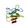
| ||||||||
|---|---|---|---|---|---|---|---|---|---|
| 1 |
| ||||||||
| Unit cell |
|
-Components
| #1: Protein | Mass: 9050.947 Da / Num. of mol.: 1 Source method: isolated from a genetically manipulated source Source: (gene. exp.) Dictyoglomus thermophilum (bacteria) / Strain: ATCC 35947 / DSM 3960 / H-6-12 / Gene: DICTH_0179Production host: References: UniProt: B5YBJ6 |
|---|---|
| #2: Water | ChemComp-HOH /  Mass: 18.015 Da / Num. of mol.: 34 / Source method: isolated from a natural source / Formula: H2O Mass: 18.015 Da / Num. of mol.: 34 / Source method: isolated from a natural source / Formula: H2O |
-Experimental details
-Experiment
| Experiment | Method: X-RAY DIFFRACTION / Number of used crystals: 1 |
|---|
- Sample preparation
| Crystal | Density Matthews: 1.96 Å3/Da / Density % sol: 37.1 % |
|---|---|
| Crystal grow | Temperature: 293.15 K / Method: vapor diffusion / pH: 7.5 Details: 0.09 M HEPES sodium salt pH 7.5, 1.26 M tri-Sodium citrate, 10 %(v/v) Glycerol |
-Data collection
| Diffraction | Mean temperature: 100 K / Serial crystal experiment: N |
|---|---|
| Diffraction source | Source: SYNCHROTRON / Site: Diamond  / Beamline: I04 / Wavelength: 0.97951 Å / Beamline: I04 / Wavelength: 0.97951 Å |
| Detector | Type: DECTRIS EIGER2 XE 16M / Detector: PIXEL / Date: Dec 9, 2019 |
| Radiation | Protocol: SINGLE WAVELENGTH / Monochromatic (M) / Laue (L): M / Scattering type: x-ray |
| Radiation wavelength | Wavelength: 0.97951 Å / Relative weight: 1 |
| Reflection | Resolution: 1.4→44.19 Å / Num. obs: 12002 / % possible obs: 86.2 % / Redundancy: 10.2 % / CC1/2: 1 / Rmerge(I) obs: 0.031 / Rpim(I) all: 0.009 / Rrim(I) all: 0.033 / Χ2: 0.87 / Net I/σ(I): 30.2 / Num. measured all: 122125 |
| Reflection shell | Resolution: 1.4→1.44 Å / % possible obs: 30 % / Redundancy: 1.7 % / Rmerge(I) obs: 0.638 / Num. measured all: 525 / Num. unique obs: 301 / CC1/2: 0.698 / Rpim(I) all: 0.579 / Rrim(I) all: 0.866 / Χ2: 0.69 / Net I/σ(I) obs: 0.8 |
- Processing
| Software |
| |||||||||||||||||||||||||||||||||||
|---|---|---|---|---|---|---|---|---|---|---|---|---|---|---|---|---|---|---|---|---|---|---|---|---|---|---|---|---|---|---|---|---|---|---|---|---|
| Refinement | Method to determine structure: MOLECULAR REPLACEMENT / Resolution: 1.41→44.19 Å / SU ML: 0.21 / Cross valid method: FREE R-VALUE / σ(F): 1.97 / Phase error: 38.27 / Stereochemistry target values: ML
| |||||||||||||||||||||||||||||||||||
| Solvent computation | Shrinkage radii: 0.9 Å / VDW probe radii: 1.11 Å / Solvent model: FLAT BULK SOLVENT MODEL | |||||||||||||||||||||||||||||||||||
| Refinement step | Cycle: LAST / Resolution: 1.41→44.19 Å
| |||||||||||||||||||||||||||||||||||
| Refine LS restraints |
| |||||||||||||||||||||||||||||||||||
| LS refinement shell |
|
