[English] 日本語
Yorodumi
- PDB-8ppz: Co-crystal structure of FKBP12, compound 7 and the FRB fragment o... -

+
Open data


ID or keywords:

Loading...

-
Basic information

Entry
Database: PDB / ID: 8ppz
TitleCo-crystal structure of FKBP12, compound 7 and the FRB fragment of mTOR
Components
  • Peptidyl-prolyl cis-trans isomerase FKBP1A
  • Serine/threonine-protein kinase mTOR
KeywordsISOMERASE / FKBP12 / mTOR / Kinase / Complex / molecular Glue
Function / homology
Function and homology information


positive regulation of cytoplasmic translational initiation / positive regulation of pentose-phosphate shunt / RNA polymerase III type 1 promoter sequence-specific DNA binding / RNA polymerase III type 2 promoter sequence-specific DNA binding / T-helper 1 cell lineage commitment / regulation of locomotor rhythm / positive regulation of wound healing, spreading of epidermal cells / cellular response to leucine starvation / regulation of membrane permeability / TFIIIC-class transcription factor complex binding ...positive regulation of cytoplasmic translational initiation / positive regulation of pentose-phosphate shunt / RNA polymerase III type 1 promoter sequence-specific DNA binding / RNA polymerase III type 2 promoter sequence-specific DNA binding / T-helper 1 cell lineage commitment / regulation of locomotor rhythm / positive regulation of wound healing, spreading of epidermal cells / cellular response to leucine starvation / regulation of membrane permeability / TFIIIC-class transcription factor complex binding / heart valve morphogenesis / negative regulation of lysosome organization / macrolide binding / RNA polymerase III type 3 promoter sequence-specific DNA binding / TORC2 complex / activin receptor binding / TORC1 complex / positive regulation of transcription of nucleolar large rRNA by RNA polymerase I / cytoplasmic side of membrane / regulation of autophagosome assembly / calcineurin-NFAT signaling cascade / nucleus localization / TORC1 signaling / voluntary musculoskeletal movement / regulation of osteoclast differentiation / positive regulation of keratinocyte migration / signaling receptor inhibitor activity / transforming growth factor beta receptor binding / TGFBR1 LBD Mutants in Cancer / cellular response to L-leucine / MTOR signalling / Amino acids regulate mTORC1 / cellular response to nutrient / energy reserve metabolic process / type I transforming growth factor beta receptor binding / Energy dependent regulation of mTOR by LKB1-AMPK / negative regulation of activin receptor signaling pathway / negative regulation of cell size / heart trabecula formation / ruffle organization / cellular response to osmotic stress / terminal cisterna / ryanodine receptor complex / I-SMAD binding / negative regulation of protein localization to nucleus / anoikis / regulation of amyloid precursor protein catabolic process / cardiac muscle cell development / positive regulation of transcription by RNA polymerase III / protein maturation by protein folding / negative regulation of calcineurin-NFAT signaling cascade / ventricular cardiac muscle tissue morphogenesis / 'de novo' protein folding / regulation of myelination / regulation of cell size / Macroautophagy / negative regulation of phosphoprotein phosphatase activity / positive regulation of oligodendrocyte differentiation / negative regulation of macroautophagy / FK506 binding / positive regulation of actin filament polymerization / lysosome organization / positive regulation of myotube differentiation / behavioral response to pain / oligodendrocyte differentiation / Constitutive Signaling by AKT1 E17K in Cancer / mTORC1-mediated signalling / TGF-beta receptor signaling activates SMADs / germ cell development / CD28 dependent PI3K/Akt signaling / cellular response to nutrient levels / positive regulation of phosphoprotein phosphatase activity / Calcineurin activates NFAT / HSF1-dependent transactivation / regulation of immune response / TOR signaling / neuronal action potential / positive regulation of translational initiation / response to amino acid / regulation of macroautophagy / endomembrane system / 'de novo' pyrimidine nucleobase biosynthetic process / protein peptidyl-prolyl isomerization / positive regulation of lamellipodium assembly / positive regulation of epithelial to mesenchymal transition / positive regulation of lipid biosynthetic process / heart morphogenesis / cardiac muscle contraction / supramolecular fiber organization / regulation of cellular response to heat / regulation of ryanodine-sensitive calcium-release channel activity / positive regulation of stress fiber assembly / cytoskeleton organization / sarcoplasmic reticulum membrane / T cell costimulation / T cell activation / cellular response to amino acid starvation / phagocytic vesicle / positive regulation of glycolytic process / cellular response to starvation
Similarity search - Function
Domain of unknown function DUF3385, target of rapamycin protein / Serine/threonine-protein kinase mTOR domain / Domain of unknown function / FKBP12-rapamycin binding domain / Serine/threonine-protein kinase TOR / FKBP12-rapamycin binding domain superfamily / FKBP12-rapamycin binding domain / Rapamycin binding domain / PIK-related kinase, FAT / FAT domain ...Domain of unknown function DUF3385, target of rapamycin protein / Serine/threonine-protein kinase mTOR domain / Domain of unknown function / FKBP12-rapamycin binding domain / Serine/threonine-protein kinase TOR / FKBP12-rapamycin binding domain superfamily / FKBP12-rapamycin binding domain / Rapamycin binding domain / PIK-related kinase, FAT / FAT domain / FATC domain / FATC / FATC domain / PIK-related kinase / FAT domain profile. / FATC domain profile. / FKBP-type peptidyl-prolyl cis-trans isomerase domain profile. / FKBP-type peptidyl-prolyl cis-trans isomerase domain / FKBP-type peptidyl-prolyl cis-trans isomerase / Phosphatidylinositol 3- and 4-kinases signature 1. / Phosphatidylinositol 3/4-kinase, conserved site / Phosphatidylinositol 3- and 4-kinases signature 2. / Phosphatidylinositol 3-/4-kinase, catalytic domain superfamily / Phosphoinositide 3-kinase, catalytic domain / Phosphatidylinositol 3- and 4-kinase / Phosphatidylinositol 3- and 4-kinases catalytic domain profile. / Phosphatidylinositol 3-/4-kinase, catalytic domain / Peptidyl-prolyl cis-trans isomerase domain superfamily / Armadillo-like helical / Tetratricopeptide-like helical domain superfamily / Armadillo-type fold / Protein kinase-like domain superfamily
Similarity search - Domain/homology
Chem-0AN / ACETATE ION / Serine/threonine-protein kinase mTOR / Peptidyl-prolyl cis-trans isomerase FKBP1A
Similarity search - Component
Biological speciesHomo sapiens (human)
MethodX-RAY DIFFRACTION / SYNCHROTRON / MOLECULAR REPLACEMENT / Resolution: 1.85 Å
AuthorsMeyners, C. / Deutscher, R.C.E. / Hausch, F.
Funding support Germany, 2items
OrganizationGrant numberCountry
German Federal Ministry for Education and Research03ZU1109EB Germany
German Research Foundation (DFG)HA5556 Germany
CitationJournal: Chemrxiv / Year: 2023
Title: Co-crystal structure of FKBP12, compound 7 and the FRB fragment of mTOR
Authors: Meyners, C. / Deutscher, R.C.E. / Hausch, F.
History
DepositionJul 10, 2023Deposition site: PDBE / Processing site: PDBE
Revision 1.0Aug 9, 2023Provider: repository / Type: Initial release
Revision 1.1Jun 19, 2024Group: Data collection / Category: chem_comp_atom / chem_comp_bond

-
Structure visualization

Structure viewerMolecule:
MolmilJmol/JSmol

Downloads & links

-
Assembly

Deposited unit
A: Peptidyl-prolyl cis-trans isomerase FKBP1A
B: Serine/threonine-protein kinase mTOR
hetero molecules


Theoretical massNumber of molelcules
Total (without water)24,3908
Polymers23,5802
Non-polymers8106
Water2,306128
1


  • Idetical with deposited unit
  • defined by author&software
TypeNameSymmetry operationNumber
identity operation1_555x,y,z1
Buried area1620 Å2
ΔGint-41 kcal/mol
Surface area10480 Å2
MethodPISA
Unit cell
Length a, b, c (Å)39.912, 64.680, 93.349
Angle α, β, γ (deg.)90.000, 90.000, 90.000
Int Tables number18
Space group name H-MP22121
Components on special symmetry positions
IDModelComponents
11B-2202-

CA

21B-2326-

HOH

31B-2331-

HOH

-
Components

-
Protein , 2 types, 2 molecules AB

#1: Protein Peptidyl-prolyl cis-trans isomerase FKBP1A / PPIase FKBP1A / 12 kDa FK506-binding protein / 12 kDa FKBP / FKBP-12 / Calstabin-1 / FK506-binding ...PPIase FKBP1A / 12 kDa FK506-binding protein / 12 kDa FKBP / FKBP-12 / Calstabin-1 / FK506-binding protein 1A / FKBP-1A / Immunophilin FKBP12 / Rotamase


Mass: 11832.496 Da / Num. of mol.: 1 / Mutation: C22V
Source method: isolated from a genetically manipulated source
Source: (gene. exp.) Homo sapiens (human) / Gene: FKBP1A, FKBP1, FKBP12
Production host: Escherichia coli 'BL21-Gold(DE3)pLysS AG' (bacteria)
References: UniProt: P62942, peptidylprolyl isomerase
#2: Protein Serine/threonine-protein kinase mTOR / FK506-binding protein 12-rapamycin complex-associated protein 1 / FKBP12-rapamycin complex- ...FK506-binding protein 12-rapamycin complex-associated protein 1 / FKBP12-rapamycin complex-associated protein / Mammalian target of rapamycin / mTOR / Mechanistic target of rapamycin / Rapamycin and FKBP12 target 1 / Rapamycin target protein 1


Mass: 11747.374 Da / Num. of mol.: 1
Source method: isolated from a genetically manipulated source
Source: (gene. exp.) Homo sapiens (human) / Gene: MTOR, FRAP, FRAP1, FRAP2, RAFT1, RAPT1
Production host: Escherichia coli 'BL21-Gold(DE3)pLysS AG' (bacteria)
References: UniProt: P42345, non-specific serine/threonine protein kinase

-
Non-polymers , 4 types, 134 molecules

#3: Chemical ChemComp-0AN / (1~{S},5~{S},6~{R})-10-[3,5-bis(chloranyl)phenyl]sulfonyl-5-[(~{E})-2-(2-chlorophenyl)ethenyl]-3-(pyridin-2-ylmethyl)-3,10-diazabicyclo[4.3.1]decan-2-one


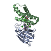
Mass: 590.948 Da / Num. of mol.: 1 / Source method: obtained synthetically / Formula: C28H26Cl3N3O3S / Feature type: SUBJECT OF INVESTIGATION
#4: Chemical
ChemComp-CA / CALCIUM ION


Mass: 40.078 Da / Num. of mol.: 4 / Source method: obtained synthetically / Formula: Ca
#5: Chemical ChemComp-ACT / ACETATE ION


Mass: 59.044 Da / Num. of mol.: 1 / Source method: obtained synthetically / Formula: C2H3O2
#6: Water ChemComp-HOH / water


Mass: 18.015 Da / Num. of mol.: 128 / Source method: isolated from a natural source / Formula: H2O

-
Details

Has ligand of interestY

-
Experimental details

-
Experiment

ExperimentMethod: X-RAY DIFFRACTION / Number of used crystals: 1

-
Sample preparation

CrystalDensity Matthews: 2.55 Å3/Da / Density % sol: 51.86 %
Crystal growTemperature: 293 K / Method: vapor diffusion, sitting drop / pH: 7.5
Details: 10% PEG8000, 0.1 M HEPES pH 7.5, 0.2 M calcium actetate

-
Data collection

DiffractionMean temperature: 100 K / Serial crystal experiment: N
Diffraction sourceSource: SYNCHROTRON / Site: PETRA III, EMBL c/o DESY / Beamline: P13 (MX1) / Wavelength: 0.97626 Å
DetectorType: DECTRIS PILATUS 6M-F / Detector: PIXEL / Date: Nov 26, 2020
RadiationProtocol: SINGLE WAVELENGTH / Monochromatic (M) / Laue (L): M / Scattering type: x-ray
Radiation wavelengthWavelength: 0.97626 Å / Relative weight: 1
ReflectionResolution: 1.85→93.35 Å / Num. obs: 20522 / % possible obs: 96 % / Redundancy: 12.8 % / CC1/2: 0.999 / Rmerge(I) obs: 0.106 / Rpim(I) all: 0.044 / Rrim(I) all: 0.115 / Net I/σ(I): 15.2
Reflection shell

Diffraction-ID: 1

Resolution (Å)Redundancy (%)Rmerge(I) obsNum. unique obsCC1/2Rpim(I) allRrim(I) all
9.06-93.3510.60.0482370.9990.0190.052
1.85-1.8912.41.36712790.8060.5791.487

-
Processing

Software
NameVersionClassification
REFMAC5.8.0415refinement
XDSdata reduction
Aimlessdata scaling
PHASERphasing
RefinementMethod to determine structure: MOLECULAR REPLACEMENT / Resolution: 1.85→46.718 Å / Cor.coef. Fo:Fc: 0.959 / Cor.coef. Fo:Fc free: 0.936 / SU B: 3.573 / SU ML: 0.101 / Cross valid method: THROUGHOUT / ESU R: 0.138 / ESU R Free: 0.137
Details: Hydrogens have been added in their riding positions
RfactorNum. reflection% reflection
Rfree0.2383 1055 5.151 %
Rwork0.192 19425 -
all0.194 --
obs-20480 96.042 %
Solvent computationIon probe radii: 0.8 Å / Shrinkage radii: 0.8 Å / VDW probe radii: 1.2 Å / Solvent model: MASK BULK SOLVENT
Displacement parametersBiso mean: 29.647 Å2
Baniso -1Baniso -2Baniso -3
1-2.918 Å20 Å2-0 Å2
2---2.693 Å20 Å2
3----0.225 Å2
Refinement stepCycle: LAST / Resolution: 1.85→46.718 Å
ProteinNucleic acidLigandSolventTotal
Num. atoms1590 0 46 128 1764
Refine LS restraints
Refine-IDTypeDev idealDev ideal targetNumber
X-RAY DIFFRACTIONr_bond_refined_d0.0090.0121675
X-RAY DIFFRACTIONr_bond_other_d0.0030.0161521
X-RAY DIFFRACTIONr_angle_refined_deg1.6611.7012263
X-RAY DIFFRACTIONr_angle_other_deg0.6381.6123503
X-RAY DIFFRACTIONr_dihedral_angle_1_deg6.2035199
X-RAY DIFFRACTIONr_dihedral_angle_2_deg6.609511
X-RAY DIFFRACTIONr_dihedral_angle_3_deg14.9710275
X-RAY DIFFRACTIONr_dihedral_angle_6_deg15.9161078
X-RAY DIFFRACTIONr_chiral_restr0.1740.2230
X-RAY DIFFRACTIONr_chiral_restr_other1.4830.25
X-RAY DIFFRACTIONr_gen_planes_refined0.0080.021980
X-RAY DIFFRACTIONr_gen_planes_other0.0010.02399
X-RAY DIFFRACTIONr_nbd_refined0.2180.2333
X-RAY DIFFRACTIONr_symmetry_nbd_other0.190.21329
X-RAY DIFFRACTIONr_nbtor_refined0.1840.2830
X-RAY DIFFRACTIONr_symmetry_nbtor_other0.080.2859
X-RAY DIFFRACTIONr_xyhbond_nbd_refined0.1680.2113
X-RAY DIFFRACTIONr_metal_ion_refined0.0840.22
X-RAY DIFFRACTIONr_symmetry_nbd_refined0.1880.212
X-RAY DIFFRACTIONr_nbd_other0.1120.241
X-RAY DIFFRACTIONr_symmetry_xyhbond_nbd_refined0.1750.225
X-RAY DIFFRACTIONr_symmetry_metal_ion_refined0.110.22
X-RAY DIFFRACTIONr_mcbond_it2.8453.113806
X-RAY DIFFRACTIONr_mcbond_other2.8213.11805
X-RAY DIFFRACTIONr_mcangle_it3.8635.5811001
X-RAY DIFFRACTIONr_mcangle_other3.8615.5841002
X-RAY DIFFRACTIONr_scbond_it3.9373.356869
X-RAY DIFFRACTIONr_scbond_other3.9353.357870
X-RAY DIFFRACTIONr_scangle_it5.73361262
X-RAY DIFFRACTIONr_scangle_other5.736.0011263
X-RAY DIFFRACTIONr_lrange_it7.46330.8811959
X-RAY DIFFRACTIONr_lrange_other7.34230.271924
LS refinement shell

Refine-ID: X-RAY DIFFRACTION / Total num. of bins used: 20

Resolution (Å)Rfactor RfreeNum. reflection RfreeRfactor RworkNum. reflection RworkRfactor allNum. reflection allFsc freeFsc work% reflection obs (%)WRfactor Rwork
1.85-1.8980.32810.29414790.29615600.9210.9061000.278
1.898-1.950.3480.3139410.31214870.9140.88766.50980.283
1.95-2.0060.307610.26412470.26614720.9240.94588.85870.239
2.006-2.0680.29900.22813290.23214190.940.9591000.202
2.068-2.1360.263760.22712930.22913690.9450.9621000.198
2.136-2.210.246630.20112800.20313430.9620.971000.176
2.21-2.2940.253570.19510630.19812950.9540.97286.48650.164
2.294-2.3870.22750.18111920.18312670.9720.9781000.157
2.387-2.4930.236620.17411410.17812030.9710.9811000.155
2.493-2.6140.217480.16911130.17111610.9760.9831000.148
2.614-2.7550.206600.16910270.1710870.9770.9821000.15
2.755-2.9220.293530.2029990.20610520.940.9731000.186
2.922-3.1230.237580.2029190.2049770.960.9741000.188
3.123-3.3710.204520.1868670.1879190.9780.9781000.178
3.371-3.6910.186340.28160.1998500.9760.9781000.194
3.691-4.1240.242370.1737450.1757870.9710.98399.36470.176
4.124-4.7560.253240.1426690.1456930.9650.9881000.151
4.756-5.8090.203320.1725700.1746030.9740.98599.83420.178
5.809-8.1530.24260.1924580.1944840.9630.981000.196
8.153-46.7180.256180.2052780.2082960.9730.9671000.215

+
About Yorodumi

-
News

-
Feb 9, 2022. New format data for meta-information of EMDB entries

New format data for meta-information of EMDB entries

  • Version 3 of the EMDB header file is now the official format.
  • The previous official version 1.9 will be removed from the archive.

Related info.:EMDB header

External links:wwPDB to switch to version 3 of the EMDB data model

-
Aug 12, 2020. Covid-19 info

Covid-19 info

URL: https://pdbjlvh1.pdbj.org/emnavi/covid19.php

New page: Covid-19 featured information page in EM Navigator.

Related info.:Covid-19 info / Mar 5, 2020. Novel coronavirus structure data

+
Mar 5, 2020. Novel coronavirus structure data

Novel coronavirus structure data

Related info.:Yorodumi Speices / Aug 12, 2020. Covid-19 info

External links:COVID-19 featured content - PDBj / Molecule of the Month (242):Coronavirus Proteases

+
Jan 31, 2019. EMDB accession codes are about to change! (news from PDBe EMDB page)

EMDB accession codes are about to change! (news from PDBe EMDB page)

  • The allocation of 4 digits for EMDB accession codes will soon come to an end. Whilst these codes will remain in use, new EMDB accession codes will include an additional digit and will expand incrementally as the available range of codes is exhausted. The current 4-digit format prefixed with “EMD-” (i.e. EMD-XXXX) will advance to a 5-digit format (i.e. EMD-XXXXX), and so on. It is currently estimated that the 4-digit codes will be depleted around Spring 2019, at which point the 5-digit format will come into force.
  • The EM Navigator/Yorodumi systems omit the EMD- prefix.

Related info.:Q: What is EMD? / ID/Accession-code notation in Yorodumi/EM Navigator

External links:EMDB Accession Codes are Changing Soon! / Contact to PDBj

+
Jul 12, 2017. Major update of PDB

Major update of PDB

  • wwPDB released updated PDB data conforming to the new PDBx/mmCIF dictionary.
  • This is a major update changing the version number from 4 to 5, and with Remediation, in which all the entries are updated.
  • In this update, many items about electron microscopy experimental information are reorganized (e.g. em_software).
  • Now, EM Navigator and Yorodumi are based on the updated data.

External links:wwPDB Remediation / Enriched Model Files Conforming to OneDep Data Standards Now Available in the PDB FTP Archive

-
Yorodumi

Thousand views of thousand structures

  • Yorodumi is a browser for structure data from EMDB, PDB, SASBDB, etc.
  • This page is also the successor to EM Navigator detail page, and also detail information page/front-end page for Omokage search.
  • The word "yorodu" (or yorozu) is an old Japanese word meaning "ten thousand". "mi" (miru) is to see.

Related info.:EMDB / PDB / SASBDB / Comparison of 3 databanks / Yorodumi Search / Aug 31, 2016. New EM Navigator & Yorodumi / Yorodumi Papers / Jmol/JSmol / Function and homology information / Changes in new EM Navigator and Yorodumi

Read more