 Movie
Movie Controller

[English] 日本語
 Yorodumi
- PDB-8ppc: Human inositol 1,4,5-trisphosphate 3-kinase A (IP3K) catalytic do... -
+ Open data

- Basic information
| Entry | Database: PDB / ID: 8ppc | |||||||||
|---|---|---|---|---|---|---|---|---|---|---|
| Title | Human inositol 1,4,5-trisphosphate 3-kinase A (IP3K) catalytic domain in complex with L-chiro-inositol 2,3,5-trisphosphate/ATP/Mn | |||||||||
 Components Components | Inositol-trisphosphate 3-kinase A | |||||||||
 Keywords Keywords | TRANSFERASE / Inositol polyphosphate / InsP / inositol kinase / IP3K / calcium / InsP3 / IP3 / IPK / IP3 3-K | |||||||||
| Function / homology |  Function and homology information Function and homology informationinositol-trisphosphate 3-kinase / inositol-1,4,5-trisphosphate 3-kinase activity / inositol hexakisphosphate kinase activity / inositol phosphate biosynthetic process / modification of postsynaptic actin cytoskeleton / inositol metabolic process / positive regulation of dendritic spine morphogenesis / postsynaptic actin cytoskeleton / calcium/calmodulin-dependent protein kinase activity / dendritic spine maintenance ...inositol-trisphosphate 3-kinase / inositol-1,4,5-trisphosphate 3-kinase activity / inositol hexakisphosphate kinase activity / inositol phosphate biosynthetic process / modification of postsynaptic actin cytoskeleton / inositol metabolic process / positive regulation of dendritic spine morphogenesis / postsynaptic actin cytoskeleton / calcium/calmodulin-dependent protein kinase activity / dendritic spine maintenance / phosphatidylinositol phosphate biosynthetic process / Synthesis of IP3 and IP4 in the cytosol / cellular response to calcium ion / response to calcium ion / regulation of synaptic plasticity / small GTPase binding / actin cytoskeleton organization / dendritic spine / cytoskeleton / calmodulin binding / glutamatergic synapse / signal transduction / ATP binding / nucleus / cytosol / cytoplasm Similarity search - Function | |||||||||
| Biological species |  Homo sapiens (human) Homo sapiens (human) | |||||||||
| Method |  X-RAY DIFFRACTION / SYNCHROTRON / FOURIER SYNTHESIS / Resolution: 1.92 Å X-RAY DIFFRACTION / SYNCHROTRON / FOURIER SYNTHESIS / Resolution: 1.92 Å | |||||||||
 Authors Authors | Marquez-Monino, M.A. / Gonzalez, B. | |||||||||
| Funding support |  Spain, 2items Spain, 2items
| |||||||||
 Citation Citation | Journal: Nat Commun / Year: 2024 Title: Substrate promiscuity of inositol 1,4,5-trisphosphate kinase driven by structurally-modified ligands and active site plasticity. Authors: Marquez-Monino, M.A. / Ortega-Garcia, R. / Whitfield, H. / Riley, A.M. / Infantes, L. / Garrett, S.W. / Shipton, M.L. / Brearley, C.A. / Potter, B.V.L. / Gonzalez, B. | |||||||||
| History |
|
- Structure visualization
| Structure viewer | Molecule: MolmilJmol/JSmol |
|---|
- Downloads & links
-Download
| PDBx/mmCIF format | 8ppc.cif.gz | 136.5 KB | Display | PDBx/mmCIF format |
|---|---|---|---|---|
| PDB format | pdb8ppc.ent.gz | 104 KB | Display | PDB format |
| PDBx/mmJSON format | 8ppc.json.gz | Tree view | PDBx/mmJSON format | |
| Others |  Other downloads Other downloads |
-Validation report
| Summary document | 8ppc_validation.pdf.gz | 2.7 MB | Display | wwPDB validaton report |
|---|---|---|---|---|
| Full document | 8ppc_full_validation.pdf.gz | 2.7 MB | Display | |
| Data in XML | 8ppc_validation.xml.gz | 24.7 KB | Display | |
| Data in CIF | 8ppc_validation.cif.gz | 35.3 KB | Display | |
| Arichive directory | https://data.pdbj.org/pub/pdb/validation_reports/pp/8ppcftp://data.pdbj.org/pub/pdb/validation_reports/pp/8ppc | HTTPS FTP |
-Related structure data
| Related structure data |  8pp8C  8pp9C  8ppaC  8ppbC  8ppdC  8ppeC  8ppfC  8ppgC  8pphC  8ppiC  8ppjC C: citing same article ( |
|---|---|
| Similar structure data |
-Links
 PDBj
PDBj


- Assembly
| Deposited unit | 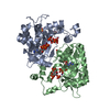
| |||||||||
|---|---|---|---|---|---|---|---|---|---|---|
| 1 | 
| |||||||||
| 2 | 
| |||||||||
| Unit cell |
| |||||||||
| Components on special symmetry positions |
|
-Components
-Protein , 1 types, 2 molecules AB
| #1: Protein | Mass: 31877.268 Da / Num. of mol.: 2 Source method: isolated from a genetically manipulated source Details: Catalytic domain / Source: (gene. exp.) Homo sapiens (human) / Gene: ITPKA / Production host:  References: UniProt: P23677, inositol-trisphosphate 3-kinase |
|---|
-Non-polymers , 5 types, 285 molecules 


| #2: Chemical | Mass: 420.096 Da / Num. of mol.: 2 / Source method: obtained synthetically / Formula: C6H15O15P3 / Feature type: SUBJECT OF INVESTIGATION #3: Chemical |  Mass: 507.181 Da / Num. of mol.: 2 / Source method: obtained synthetically / Formula: C10H16N5O13P3 / Feature type: SUBJECT OF INVESTIGATION / Comment: ATP, energy-carrying molecule*YM Mass: 507.181 Da / Num. of mol.: 2 / Source method: obtained synthetically / Formula: C10H16N5O13P3 / Feature type: SUBJECT OF INVESTIGATION / Comment: ATP, energy-carrying molecule*YM#4: Chemical |  Mass: 54.938 Da / Num. of mol.: 2 / Source method: obtained synthetically / Formula: Mn / Feature type: SUBJECT OF INVESTIGATION Mass: 54.938 Da / Num. of mol.: 2 / Source method: obtained synthetically / Formula: Mn / Feature type: SUBJECT OF INVESTIGATION#5: Chemical | ChemComp-SO4 /  Mass: 96.063 Da / Num. of mol.: 6 / Source method: obtained synthetically / Formula: SO4 Mass: 96.063 Da / Num. of mol.: 6 / Source method: obtained synthetically / Formula: SO4#6: Water | ChemComp-HOH / | Mass: 18.015 Da / Num. of mol.: 273 / Source method: isolated from a natural source / Formula: H2O |
|---|
-Details
| Has ligand of interest | Y |
|---|
-Experimental details
-Experiment
| Experiment | Method: X-RAY DIFFRACTION / Number of used crystals: 1 |
|---|
- Sample preparation
| Crystal | Density Matthews: 2.69 Å3/Da / Density % sol: 54.29 % / Description: Cube / Trapezoidal |
|---|---|
| Crystal grow | Temperature: 291 K / Method: vapor diffusion, sitting drop / pH: 8.5 Details: 0.80 M sodium citrate, 0.1M Tris pH 8.5 and 0.1 M NaCl. Protein:precipitant ratio 1:1. Protein concentration: 9 mg/ml. Protein buffer: 20 mM Tris pH 7.5, 50 mM ammonium sulfate and 2 mM DTT. ...Details: 0.80 M sodium citrate, 0.1M Tris pH 8.5 and 0.1 M NaCl. Protein:precipitant ratio 1:1. Protein concentration: 9 mg/ml. Protein buffer: 20 mM Tris pH 7.5, 50 mM ammonium sulfate and 2 mM DTT. Soaking 2h with 1.5 M lithium sulfate, 0.1 M Tris pH 8.5, 5 mM ligand, 3 mM ATP and 3 mM MnCl2. |
-Data collection
| Diffraction | Mean temperature: 100 K / Serial crystal experiment: N |
|---|---|
| Diffraction source | Source: SYNCHROTRON / Site: ALBA / Beamline: XALOC / Wavelength: 0.979264 Å |
| Detector | Type: DECTRIS PILATUS 6M / Detector: PIXEL / Date: Jun 19, 2021 / Details: KB mirrors, rectangular beam shape |
| Radiation | Monochromator: Channel-cut Si(111) / Protocol: SINGLE WAVELENGTH / Monochromatic (M) / Laue (L): M / Scattering type: x-ray |
| Radiation wavelength | Wavelength: 0.979264 Å / Relative weight: 1 |
| Reflection | Resolution: 1.92→49.66 Å / Num. obs: 51230 / % possible obs: 98.4 % / Redundancy: 10 % / Biso Wilson estimate: 31.616 Å2 / CC1/2: 0.997 / Rpim(I) all: 0.032 / Net I/σ(I): 13.3 |
| Reflection shell | Resolution: 1.92→1.97 Å / Redundancy: 7.6 % / Mean I/σ(I) obs: 1.8 / Num. unique obs: 2906 / CC1/2: 0.799 / Rpim(I) all: 0.299 / % possible all: 84.8 |
- Processing
| Software |
| ||||||||||||||||||||||||||||||||||||||||||||||||||||||||||||||||||||||||||||||||||||||||||||||||||||||||||||||||||||||||||||||||||||||||||||||||||||||||||||||||||||||||||||||||||||||
|---|---|---|---|---|---|---|---|---|---|---|---|---|---|---|---|---|---|---|---|---|---|---|---|---|---|---|---|---|---|---|---|---|---|---|---|---|---|---|---|---|---|---|---|---|---|---|---|---|---|---|---|---|---|---|---|---|---|---|---|---|---|---|---|---|---|---|---|---|---|---|---|---|---|---|---|---|---|---|---|---|---|---|---|---|---|---|---|---|---|---|---|---|---|---|---|---|---|---|---|---|---|---|---|---|---|---|---|---|---|---|---|---|---|---|---|---|---|---|---|---|---|---|---|---|---|---|---|---|---|---|---|---|---|---|---|---|---|---|---|---|---|---|---|---|---|---|---|---|---|---|---|---|---|---|---|---|---|---|---|---|---|---|---|---|---|---|---|---|---|---|---|---|---|---|---|---|---|---|---|---|---|---|---|
| Refinement | Method to determine structure: FOURIER SYNTHESIS / Resolution: 1.92→49.66 Å / Cor.coef. Fo:Fc: 0.96 / Cor.coef. Fo:Fc free: 0.946 / SU B: 3.385 / SU ML: 0.095 / Cross valid method: THROUGHOUT / ESU R: 0.156 / ESU R Free: 0.137 / Stereochemistry target values: MAXIMUM LIKELIHOOD / Details: HYDROGENS HAVE BEEN ADDED IN THE RIDING POSITIONS
| ||||||||||||||||||||||||||||||||||||||||||||||||||||||||||||||||||||||||||||||||||||||||||||||||||||||||||||||||||||||||||||||||||||||||||||||||||||||||||||||||||||||||||||||||||||||
| Solvent computation | Ion probe radii: 0.8 Å / Shrinkage radii: 0.8 Å / VDW probe radii: 1.2 Å / Solvent model: MASK | ||||||||||||||||||||||||||||||||||||||||||||||||||||||||||||||||||||||||||||||||||||||||||||||||||||||||||||||||||||||||||||||||||||||||||||||||||||||||||||||||||||||||||||||||||||||
| Displacement parameters | Biso mean: 42.024 Å2
| ||||||||||||||||||||||||||||||||||||||||||||||||||||||||||||||||||||||||||||||||||||||||||||||||||||||||||||||||||||||||||||||||||||||||||||||||||||||||||||||||||||||||||||||||||||||
| Refinement step | Cycle: 1 / Resolution: 1.92→49.66 Å
| ||||||||||||||||||||||||||||||||||||||||||||||||||||||||||||||||||||||||||||||||||||||||||||||||||||||||||||||||||||||||||||||||||||||||||||||||||||||||||||||||||||||||||||||||||||||
| Refine LS restraints |
|