 Movie
Movie Controller


+ Open data

- Basic information
| Entry | Database: PDB / ID: 8pnb | |||||||||||||||||||||||||||||||||
|---|---|---|---|---|---|---|---|---|---|---|---|---|---|---|---|---|---|---|---|---|---|---|---|---|---|---|---|---|---|---|---|---|---|---|
| Title | HRV empty capsid | |||||||||||||||||||||||||||||||||
 Components Components |
| |||||||||||||||||||||||||||||||||
 Keywords Keywords | VIRUS / cryo-EM / HRV-B14 / SsRNA virus / capsid | |||||||||||||||||||||||||||||||||
| Function / homology |  Function and homology information Function and homology informationlysis of host organelle involved in viral entry into host cell / symbiont-mediated suppression of host cytoplasmic pattern recognition receptor signaling pathway via inhibition of RIG-I activity / picornain 2A / symbiont-mediated suppression of host mRNA export from nucleus / symbiont genome entry into host cell via pore formation in plasma membrane / picornain 3C / T=pseudo3 icosahedral viral capsid / host cell cytoplasmic vesicle membrane / ribonucleoside triphosphate phosphatase activity / nucleoside-triphosphate phosphatase ...lysis of host organelle involved in viral entry into host cell / symbiont-mediated suppression of host cytoplasmic pattern recognition receptor signaling pathway via inhibition of RIG-I activity / picornain 2A / symbiont-mediated suppression of host mRNA export from nucleus / symbiont genome entry into host cell via pore formation in plasma membrane / picornain 3C / T=pseudo3 icosahedral viral capsid / host cell cytoplasmic vesicle membrane / ribonucleoside triphosphate phosphatase activity / nucleoside-triphosphate phosphatase / channel activity / monoatomic ion transmembrane transport / DNA replication / RNA helicase activity / endocytosis involved in viral entry into host cell / symbiont-mediated activation of host autophagy / RNA-directed RNA polymerase / cysteine-type endopeptidase activity / viral RNA genome replication / RNA-directed RNA polymerase activity / virion attachment to host cell / host cell nucleus / structural molecule activity / DNA-templated transcription / proteolysis / RNA binding / zinc ion binding / ATP binding Similarity search - Function | |||||||||||||||||||||||||||||||||
| Biological species |  rhinovirus B14 rhinovirus B14 | |||||||||||||||||||||||||||||||||
| Method | ELECTRON MICROSCOPY / single particle reconstruction / cryo EM / Resolution: 3.8 Å | |||||||||||||||||||||||||||||||||
 Authors Authors | Gil-Cantero, D. / Mata, C.P. / Mateu, M.G. / Caston, J.R. | |||||||||||||||||||||||||||||||||
| Funding support |  Spain, 1items Spain, 1items
| |||||||||||||||||||||||||||||||||
 Citation Citation | Journal: Commun Biol / Year: 2024 Title: Cryo-EM of human rhinovirus reveals capsid-RNA duplex interactions that provide insights into virus assembly and genome uncoating. Authors: David Gil-Cantero / Carlos P Mata / Luis Valiente / Alicia Rodríguez-Huete / Alejandro Valbuena / Reidun Twarock / Peter G Stockley / Mauricio G Mateu / José R Castón /  Abstract: The cryo-EM structure of the human rhinovirus B14 determined in this study reveals 13-bp RNA duplexes symmetrically bound to regions around each of the 30 two-fold axes in the icosahedral viral ...The cryo-EM structure of the human rhinovirus B14 determined in this study reveals 13-bp RNA duplexes symmetrically bound to regions around each of the 30 two-fold axes in the icosahedral viral capsid. The RNA duplexes (~12% of the ssRNA genome) define a quasi-dodecahedral cage that line a substantial part of the capsid interior surface. The RNA duplexes establish a complex network of non-covalent interactions with pockets in the capsid inner wall, including coulombic interactions with a cluster of basic amino acid residues that surround each RNA duplex. A direct comparison was made between the cryo-EM structure of RNA-filled virions and that of RNA-free (empty) capsids that resulted from genome release from a small fraction of viruses. The comparison reveals that some specific residues involved in capsid-duplex RNA interactions in the virion undergo remarkable conformational rearrangements upon RNA release from the capsid. RNA release is also associated with the asynchronous opening of channels at the 30 two-fold axes. The results provide further insights into the molecular mechanisms leading to assembly of rhinovirus particles and their genome uncoating during infection. They may also contribute to development of novel antiviral strategies aimed at interfering with viral capsid-genome interactions during the infectious cycle. | |||||||||||||||||||||||||||||||||
| History |
|
- Structure visualization
| Structure viewer | Molecule: MolmilJmol/JSmol |
|---|
- Downloads & links
-Download
| PDBx/mmCIF format | 8pnb.cif.gz | 141.7 KB | Display | PDBx/mmCIF format |
|---|---|---|---|---|
| PDB format | pdb8pnb.ent.gz | Display | PDB format | |
| PDBx/mmJSON format | 8pnb.json.gz | Tree view | PDBx/mmJSON format | |
| Others |  Other downloads Other downloads |
-Validation report
| Arichive directory | https://data.pdbj.org/pub/pdb/validation_reports/pn/8pnbftp://data.pdbj.org/pub/pdb/validation_reports/pn/8pnb | HTTPS FTP |
|---|
-Related structure data
| Related structure data |  17780MC 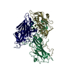 8pnfC M: map data used to model this data C: citing same article ( |
|---|---|
| Similar structure data |

-Links
 PDBj
PDBj

- Assembly
| Deposited unit | 
|
|---|---|
| 1 | x 60
|
-Components
| #1: Protein | Mass: 26233.420 Da / Num. of mol.: 1 Source method: isolated from a genetically manipulated source Source: (gene. exp.) rhinovirus B14 / Production host: rhinovirus B14 / References: UniProt: P03303 |
|---|---|
| #2: Protein | Mass: 27903.746 Da / Num. of mol.: 1 Source method: isolated from a genetically manipulated source Source: (gene. exp.) rhinovirus B14 / Production host: rhinovirus B14 / References: UniProt: P03303 |
| #3: Protein | Mass: 26236.754 Da / Num. of mol.: 1 Source method: isolated from a genetically manipulated source Source: (gene. exp.) rhinovirus B14 / Production host: rhinovirus B14References: UniProt: P03303, picornain 2A, nucleoside-triphosphate phosphatase, picornain 3C, RNA-directed RNA polymerase |
| Has protein modification | N |
-Experimental details
-Experiment
| Experiment | Method: ELECTRON MICROSCOPY |
|---|---|
| EM experiment | Aggregation state: PARTICLE / 3D reconstruction method: single particle reconstruction |
- Sample preparation
| Component | Name: rhinovirus B14 / Type: VIRUS / Details: Purified from infected H1 HeLa / Entity ID: all / Source: RECOMBINANT | |||||||||||||||||||||||||
|---|---|---|---|---|---|---|---|---|---|---|---|---|---|---|---|---|---|---|---|---|---|---|---|---|---|---|
| Molecular weight | Units: KILODALTONS/NANOMETER / Experimental value: NO | |||||||||||||||||||||||||
| Source (natural) | Organism: rhinovirus B14 | |||||||||||||||||||||||||
| Source (recombinant) | Organism: rhinovirus B14 | |||||||||||||||||||||||||
| Details of virus | Empty: YES / Enveloped: NO / Isolate: SPECIES / Type: VIRION | |||||||||||||||||||||||||
| Natural host | Organism: Homo sapiens | |||||||||||||||||||||||||
| Virus shell | Diameter: 300 nm / Triangulation number (T number): 3 | |||||||||||||||||||||||||
| Buffer solution | pH: 7.4 | |||||||||||||||||||||||||
| Buffer component |
| |||||||||||||||||||||||||
| Specimen | Embedding applied: NO / Shadowing applied: NO / Staining applied: NO / Vitrification applied: YES | |||||||||||||||||||||||||
| Specimen support | Grid material: COPPER/RHODIUM / Grid mesh size: 300 divisions/in. / Grid type: Quantifoil R2/2 | |||||||||||||||||||||||||
| Vitrification | Instrument: FEI VITROBOT MARK IV / Cryogen name: ETHANE / Humidity: 95 % / Chamber temperature: 22 K |
- Electron microscopy imaging
| Microscopy | Model: TFS TALOS |
|---|---|
| Electron gun | Electron source:  FIELD EMISSION GUN / Accelerating voltage: 200 kV / Illumination mode: OTHER FIELD EMISSION GUN / Accelerating voltage: 200 kV / Illumination mode: OTHER |
| Electron lens | Mode: OTHER / Nominal magnification: 73000 X / Nominal defocus max: 3200 nm / Nominal defocus min: 1200 nm / Cs: 2.7 mm |
| Specimen holder | Cryogen: NITROGEN |
| Image recording | Electron dose: 30 e/Å2 / Detector mode: INTEGRATING / Film or detector model: FEI FALCON III (4k x 4k) |
- Processing
| EM software |
| ||||||||||||||||||||||||
|---|---|---|---|---|---|---|---|---|---|---|---|---|---|---|---|---|---|---|---|---|---|---|---|---|---|
| CTF correction | Type: PHASE FLIPPING ONLY | ||||||||||||||||||||||||
| 3D reconstruction | Resolution: 3.8 Å / Resolution method: FSC 0.143 CUT-OFF / Num. of particles: 8381 / Algorithm: FOURIER SPACE / Num. of class averages: 1 / Symmetry type: POINT | ||||||||||||||||||||||||
| Refine LS restraints |
|
