 Movie
Movie Controller

[English] 日本語
 Yorodumi
- PDB-8pkr: Crystal structure of PknD-345, a repeat fragment from the M. tube... -
+ Open data

- Basic information
| Entry | Database: PDB / ID: 8pkr | ||||||||||||
|---|---|---|---|---|---|---|---|---|---|---|---|---|---|
| Title | Crystal structure of PknD-345, a repeat fragment from the M. tuberculosis PknD beta-propeller | ||||||||||||
 Components Components | non-specific serine/threonine protein kinase | ||||||||||||
 Keywords Keywords | UNKNOWN FUNCTION / fragment / scaffold / beta propeller / globular / PknD | ||||||||||||
| Function / homology |  Function and homology information Function and homology informationregulation of primary metabolic process / non-specific serine/threonine protein kinase / protein serine/threonine kinase activity / ATP binding / plasma membrane Similarity search - Function | ||||||||||||
| Biological species |   Mycobacterium tuberculosis (bacteria) Mycobacterium tuberculosis (bacteria) | ||||||||||||
| Method |  X-RAY DIFFRACTION / SYNCHROTRON / MOLECULAR REPLACEMENT / Resolution: 0.95 Å X-RAY DIFFRACTION / SYNCHROTRON / MOLECULAR REPLACEMENT / Resolution: 0.95 Å | ||||||||||||
 Authors Authors | Wouters, S.M.L. | ||||||||||||
| Funding support |  Belgium, 3items Belgium, 3items
| ||||||||||||
 Citation Citation | Journal: To Be Published Title: Computational design of the SAKe scaffold proteins Authors: Wouters, S.M.L. / Noguchi, H. / Voet, A.R.D. | ||||||||||||
| History |
|
- Structure visualization
| Structure viewer | Molecule: MolmilJmol/JSmol |
|---|
- Downloads & links
-Download
| PDBx/mmCIF format | 8pkr.cif.gz | 164.5 KB | Display | PDBx/mmCIF format |
|---|---|---|---|---|
| PDB format | pdb8pkr.ent.gz | 126.2 KB | Display | PDB format |
| PDBx/mmJSON format | 8pkr.json.gz | Tree view | PDBx/mmJSON format | |
| Others |  Other downloads Other downloads |
-Validation report
| Arichive directory | https://data.pdbj.org/pub/pdb/validation_reports/pk/8pkrftp://data.pdbj.org/pub/pdb/validation_reports/pk/8pkr | HTTPS FTP |
|---|
-Related structure data
| Related structure data |  8pjqC  8pjsC  8pjtC  8pjuC  8pjvC 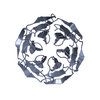 8pjwC  8pjxC  8pjyC 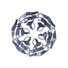 8pjzC C: citing same article ( |
|---|---|
| Similar structure data |
-Links
 PDBj
PDBj


- Assembly
| Deposited unit | 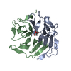
| ||||||||||
|---|---|---|---|---|---|---|---|---|---|---|---|
| 1 |
| ||||||||||
| Unit cell |
|
-Components
| #1: Protein | Mass: 13638.945 Da / Num. of mol.: 2 Source method: isolated from a genetically manipulated source Source: (gene. exp.) Mycobacterium tuberculosis (bacteria)Gene: pknD_1, pknD_2, ERS007657_00042, ERS007663_00011, ERS007665_00696, ERS007720_00884, SAMEA2683035_02262 Production host: References: UniProt: A0A045HV41, non-specific serine/threonine protein kinase #2: Chemical | ChemComp-GOL / |   Mass: 92.094 Da / Num. of mol.: 1 / Source method: obtained synthetically / Formula: C3H8O3 Mass: 92.094 Da / Num. of mol.: 1 / Source method: obtained synthetically / Formula: C3H8O3#3: Water | ChemComp-HOH / |  Mass: 18.015 Da / Num. of mol.: 235 / Source method: isolated from a natural source / Formula: H2O Mass: 18.015 Da / Num. of mol.: 235 / Source method: isolated from a natural source / Formula: H2OHas ligand of interest | N | |
|---|
-Experimental details
-Experiment
| Experiment | Method: X-RAY DIFFRACTION / Number of used crystals: 1 |
|---|
- Sample preparation
| Crystal | Density Matthews: 2.08 Å3/Da / Density % sol: 40.92 % |
|---|---|
| Crystal grow | Temperature: 293.15 K / Method: vapor diffusion Details: 0.02 M Calcium chloride, 0.1 M Sodium acetate pH 4.6, 26% (v/v) MPD |
-Data collection
| Diffraction | Mean temperature: 100 K / Serial crystal experiment: N |
|---|---|
| Diffraction source | Source: SYNCHROTRON / Site: Diamond  / Beamline: I24 / Wavelength: 0.74999 Å / Beamline: I24 / Wavelength: 0.74999 Å |
| Detector | Type: DECTRIS PILATUS3 6M / Detector: PIXEL / Date: Feb 22, 2017 |
| Radiation | Protocol: SINGLE WAVELENGTH / Monochromatic (M) / Laue (L): M / Scattering type: x-ray |
| Radiation wavelength | Wavelength: 0.74999 Å / Relative weight: 1 |
| Reflection | Resolution: 0.95→41.73 Å / Num. obs: 142745 / % possible obs: 99.1 % / Redundancy: 13.1 % / Biso Wilson estimate: 8.87 Å2 / CC1/2: 1 / Rmerge(I) obs: 0.043 / Rpim(I) all: 0.012 / Rrim(I) all: 0.045 / Net I/σ(I): 26.9 / Num. measured all: 1871882 |
| Reflection shell | Resolution: 0.95→0.97 Å / % possible obs: 97.7 % / Redundancy: 13.4 % / Rmerge(I) obs: 1.223 / Num. measured all: 92378 / Num. unique obs: 6894 / CC1/2: 0.813 / Rpim(I) all: 0.342 / Rrim(I) all: 1.271 / Net I/σ(I) obs: 2.3 |
- Processing
| Software |
| |||||||||||||||||||||||||||||||||||||||||||||||||||||||||||||||||||||||||||||||||||||||||||||||||||||||||||||||||||||||||||||||||||||||||||||||||||||||||||||||||||||||||||||||||||||||||||||||||||||||||||||||||||||||||
|---|---|---|---|---|---|---|---|---|---|---|---|---|---|---|---|---|---|---|---|---|---|---|---|---|---|---|---|---|---|---|---|---|---|---|---|---|---|---|---|---|---|---|---|---|---|---|---|---|---|---|---|---|---|---|---|---|---|---|---|---|---|---|---|---|---|---|---|---|---|---|---|---|---|---|---|---|---|---|---|---|---|---|---|---|---|---|---|---|---|---|---|---|---|---|---|---|---|---|---|---|---|---|---|---|---|---|---|---|---|---|---|---|---|---|---|---|---|---|---|---|---|---|---|---|---|---|---|---|---|---|---|---|---|---|---|---|---|---|---|---|---|---|---|---|---|---|---|---|---|---|---|---|---|---|---|---|---|---|---|---|---|---|---|---|---|---|---|---|---|---|---|---|---|---|---|---|---|---|---|---|---|---|---|---|---|---|---|---|---|---|---|---|---|---|---|---|---|---|---|---|---|---|---|---|---|---|---|---|---|---|---|---|---|---|---|---|---|---|
| Refinement | Method to determine structure: MOLECULAR REPLACEMENT / Resolution: 0.95→30.45 Å / SU ML: 0.0724 / Cross valid method: FREE R-VALUE / σ(F): 1.34 / Phase error: 10.8557 Stereochemistry target values: GeoStd + Monomer Library + CDL v1.2
| |||||||||||||||||||||||||||||||||||||||||||||||||||||||||||||||||||||||||||||||||||||||||||||||||||||||||||||||||||||||||||||||||||||||||||||||||||||||||||||||||||||||||||||||||||||||||||||||||||||||||||||||||||||||||
| Solvent computation | Shrinkage radii: 0.9 Å / VDW probe radii: 1.11 Å / Solvent model: FLAT BULK SOLVENT MODEL | |||||||||||||||||||||||||||||||||||||||||||||||||||||||||||||||||||||||||||||||||||||||||||||||||||||||||||||||||||||||||||||||||||||||||||||||||||||||||||||||||||||||||||||||||||||||||||||||||||||||||||||||||||||||||
| Displacement parameters | Biso mean: 11.86 Å2 | |||||||||||||||||||||||||||||||||||||||||||||||||||||||||||||||||||||||||||||||||||||||||||||||||||||||||||||||||||||||||||||||||||||||||||||||||||||||||||||||||||||||||||||||||||||||||||||||||||||||||||||||||||||||||
| Refinement step | Cycle: LAST / Resolution: 0.95→30.45 Å
| |||||||||||||||||||||||||||||||||||||||||||||||||||||||||||||||||||||||||||||||||||||||||||||||||||||||||||||||||||||||||||||||||||||||||||||||||||||||||||||||||||||||||||||||||||||||||||||||||||||||||||||||||||||||||
| Refine LS restraints |
| |||||||||||||||||||||||||||||||||||||||||||||||||||||||||||||||||||||||||||||||||||||||||||||||||||||||||||||||||||||||||||||||||||||||||||||||||||||||||||||||||||||||||||||||||||||||||||||||||||||||||||||||||||||||||
| LS refinement shell |
|
