 Movie
Movie Controller

[English] 日本語
 Yorodumi
- PDB-8pir: Crystal structure of Ser33 in complex with 3-PGA (3-phosphoglycerate) -
+ Open data

- Basic information
| Entry | Database: PDB / ID: 8pir | ||||||
|---|---|---|---|---|---|---|---|
| Title | Crystal structure of Ser33 in complex with 3-PGA (3-phosphoglycerate) | ||||||
 Components Components | phosphoglycerate dehydrogenase | ||||||
 Keywords Keywords | CYTOSOLIC PROTEIN / Enzyme Protein | ||||||
| Function / homology | 3-PHOSPHOGLYCERIC ACID / 1,4-BUTANEDIOL / NICOTINAMIDE-ADENINE-DINUCLEOTIDE / :  Function and homology information Function and homology information | ||||||
| Biological species |  | ||||||
| Method |  X-RAY DIFFRACTION / SYNCHROTRON / MOLECULAR REPLACEMENT / Resolution: 2.7 Å X-RAY DIFFRACTION / SYNCHROTRON / MOLECULAR REPLACEMENT / Resolution: 2.7 Å | ||||||
 Authors Authors | Perrone, S. / Cifuente, J.O. / Marina, A. / Mastrella, L. / Trastoy, B. / Linster, C.L. / Guerin, M.E. | ||||||
| Funding support |  Spain, 1items Spain, 1items
| ||||||
 Citation Citation | Journal: To Be Published Title: Crystal structure of Ser33 in complex with 3-PGA (3-phosphoglycerate) Authors: Perrone, S. / Cifuente, J.O. / Marina, A. / Mastrella, L. / Trastoy, B. / Linster, C.L. / Guerin, M.E. | ||||||
| History |
|
- Structure visualization
| Structure viewer | Molecule: MolmilJmol/JSmol |
|---|
- Downloads & links
-Download
| PDBx/mmCIF format | 8pir.cif.gz | 335.8 KB | Display | PDBx/mmCIF format |
|---|---|---|---|---|
| PDB format | pdb8pir.ent.gz | 271.4 KB | Display | PDB format |
| PDBx/mmJSON format | 8pir.json.gz | Tree view | PDBx/mmJSON format | |
| Others |  Other downloads Other downloads |
-Validation report
| Summary document | 8pir_validation.pdf.gz | 3.5 MB | Display | wwPDB validaton report |
|---|---|---|---|---|
| Full document | 8pir_full_validation.pdf.gz | 3.6 MB | Display | |
| Data in XML | 8pir_validation.xml.gz | 71 KB | Display | |
| Data in CIF | 8pir_validation.cif.gz | 89.3 KB | Display | |
| Arichive directory | https://data.pdbj.org/pub/pdb/validation_reports/pi/8pirftp://data.pdbj.org/pub/pdb/validation_reports/pi/8pir | HTTPS FTP |
-Related structure data
| Similar structure data |
|---|
-Links
 PDBj
PDBj


- Assembly
| Deposited unit | 
| ||||||||
|---|---|---|---|---|---|---|---|---|---|
| 1 |
| ||||||||
| Unit cell |
|
-Components
| #1: Protein | Mass: 51515.289 Da / Num. of mol.: 4 Source method: isolated from a genetically manipulated source Source: (gene. exp.) Gene: SER33 / Production host:  #2: Chemical | ChemComp-NAD /   Mass: 663.425 Da / Num. of mol.: 4 / Source method: obtained synthetically / Formula: C21H27N7O14P2 / Feature type: SUBJECT OF INVESTIGATION / Comment: NAD*YM Mass: 663.425 Da / Num. of mol.: 4 / Source method: obtained synthetically / Formula: C21H27N7O14P2 / Feature type: SUBJECT OF INVESTIGATION / Comment: NAD*YM#3: Chemical | ChemComp-3PG /   Mass: 186.057 Da / Num. of mol.: 4 / Source method: obtained synthetically / Formula: C3H7O7P / Feature type: SUBJECT OF INVESTIGATION Mass: 186.057 Da / Num. of mol.: 4 / Source method: obtained synthetically / Formula: C3H7O7P / Feature type: SUBJECT OF INVESTIGATION#4: Chemical | ChemComp-BU1 / 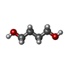  Mass: 90.121 Da / Num. of mol.: 4 / Source method: obtained synthetically / Formula: C4H10O2 / Feature type: SUBJECT OF INVESTIGATION Mass: 90.121 Da / Num. of mol.: 4 / Source method: obtained synthetically / Formula: C4H10O2 / Feature type: SUBJECT OF INVESTIGATION#5: Water | ChemComp-HOH / |  Mass: 18.015 Da / Num. of mol.: 35 / Source method: isolated from a natural source / Formula: H2O Mass: 18.015 Da / Num. of mol.: 35 / Source method: isolated from a natural source / Formula: H2OHas ligand of interest | Y | Has protein modification | N | |
|---|
-Experimental details
-Experiment
| Experiment | Method: X-RAY DIFFRACTION / Number of used crystals: 1 |
|---|
- Sample preparation
| Crystal | Density Matthews: 2.9 Å3/Da / Density % sol: 57.54 % |
|---|---|
| Crystal grow | Temperature: 291 K / Method: vapor diffusion, sitting drop Details: 0.2M 1,6-Hexanediol; 0.2M 1-Butanol; 0.2M 1,2-Propanediol; 0.2M 2-Propanol; 0.2M 1,4-Butanediol; 0.2M 1,3-Propanediol 0.1M Sodium HEPES; MOPS (acid) pH=7.5 20% v/v Glycerol; 10% w/v PEG 4000 ...Details: 0.2M 1,6-Hexanediol; 0.2M 1-Butanol; 0.2M 1,2-Propanediol; 0.2M 2-Propanol; 0.2M 1,4-Butanediol; 0.2M 1,3-Propanediol 0.1M Sodium HEPES; MOPS (acid) pH=7.5 20% v/v Glycerol; 10% w/v PEG 4000 7.4 mg/ml of protein in 25 mM Tris pH=7.5 and 150 mM NaCl |
-Data collection
| Diffraction | Mean temperature: 100 K / Serial crystal experiment: N |
|---|---|
| Diffraction source | Source: SYNCHROTRON / Site: ALBA / Beamline: XALOC / Wavelength: 0.9793 Å |
| Detector | Type: DECTRIS PILATUS 6M / Detector: PIXEL / Date: Feb 26, 2021 |
| Radiation | Protocol: SINGLE WAVELENGTH / Monochromatic (M) / Laue (L): M / Scattering type: x-ray |
| Radiation wavelength | Wavelength: 0.9793 Å / Relative weight: 1 |
| Reflection | Resolution: 2.7→60.45 Å / Num. obs: 61940 / % possible obs: 97.73 % / Redundancy: 3.5 % / CC1/2: 0.998 / Net I/σ(I): 13.01 |
| Reflection shell | Resolution: 2.7→2.797 Å / Mean I/σ(I) obs: 2.61 / Num. unique obs: 6232 / CC1/2: 0.905 |
- Processing
| Software |
| |||||||||||||||||||||||||||||||||||||||||||||||||||||||||||||||||||||||||||||||||||||||||||||||||||||||||||||||||||||||||||||||||||||||||||||||||||||||||||||||||
|---|---|---|---|---|---|---|---|---|---|---|---|---|---|---|---|---|---|---|---|---|---|---|---|---|---|---|---|---|---|---|---|---|---|---|---|---|---|---|---|---|---|---|---|---|---|---|---|---|---|---|---|---|---|---|---|---|---|---|---|---|---|---|---|---|---|---|---|---|---|---|---|---|---|---|---|---|---|---|---|---|---|---|---|---|---|---|---|---|---|---|---|---|---|---|---|---|---|---|---|---|---|---|---|---|---|---|---|---|---|---|---|---|---|---|---|---|---|---|---|---|---|---|---|---|---|---|---|---|---|---|---|---|---|---|---|---|---|---|---|---|---|---|---|---|---|---|---|---|---|---|---|---|---|---|---|---|---|---|---|---|---|---|
| Refinement | Method to determine structure: MOLECULAR REPLACEMENT / Resolution: 2.7→60.45 Å / SU ML: 0.36 / Cross valid method: FREE R-VALUE / σ(F): 1.98 / Phase error: 30.18 / Stereochemistry target values: ML
| |||||||||||||||||||||||||||||||||||||||||||||||||||||||||||||||||||||||||||||||||||||||||||||||||||||||||||||||||||||||||||||||||||||||||||||||||||||||||||||||||
| Solvent computation | Shrinkage radii: 0.9 Å / VDW probe radii: 1.11 Å / Solvent model: FLAT BULK SOLVENT MODEL | |||||||||||||||||||||||||||||||||||||||||||||||||||||||||||||||||||||||||||||||||||||||||||||||||||||||||||||||||||||||||||||||||||||||||||||||||||||||||||||||||
| Refinement step | Cycle: LAST / Resolution: 2.7→60.45 Å
| |||||||||||||||||||||||||||||||||||||||||||||||||||||||||||||||||||||||||||||||||||||||||||||||||||||||||||||||||||||||||||||||||||||||||||||||||||||||||||||||||
| Refine LS restraints |
| |||||||||||||||||||||||||||||||||||||||||||||||||||||||||||||||||||||||||||||||||||||||||||||||||||||||||||||||||||||||||||||||||||||||||||||||||||||||||||||||||
| LS refinement shell |
|
