 Movie
Movie Controller

[English] 日本語
 Yorodumi
- PDB-8pfh: Crystal structure of the yeast septin complex Shs1-Cdc12-Cdc3-Cdc10 -
+ Open data

- Basic information
| Entry | Database: PDB / ID: 8pfh | ||||||
|---|---|---|---|---|---|---|---|
| Title | Crystal structure of the yeast septin complex Shs1-Cdc12-Cdc3-Cdc10 | ||||||
 Components Components |
| ||||||
 Keywords Keywords | STRUCTURAL PROTEIN / Septins / complex | ||||||
| Function / homology |  Function and homology information Function and homology informationseptin ring disassembly / septin filament array / cellular bud neck septin ring organization / Gin4 complex / mating projection base / ascospore wall / cellular bud neck septin ring / cell cycle / septin complex / septin ring assembly ...septin ring disassembly / septin filament array / cellular bud neck septin ring organization / Gin4 complex / mating projection base / ascospore wall / cellular bud neck septin ring / cell cycle / septin complex / septin ring assembly / prospore membrane / septum digestion after cytokinesis / phosphatidylinositol-5-phosphate binding / septin ring / cytoskeleton-dependent cytokinesis / cellular bud neck / phosphatidylinositol-4-phosphate binding / cell division site / mitotic cytokinesis / structural constituent of cytoskeleton / intracellular protein localization / microtubule cytoskeleton / molecular adaptor activity / GTPase activity / GTP binding Similarity search - Function | ||||||
| Biological species |  | ||||||
| Method |  X-RAY DIFFRACTION / SYNCHROTRON / MOLECULAR REPLACEMENT / Resolution: 3.24 Å X-RAY DIFFRACTION / SYNCHROTRON / MOLECULAR REPLACEMENT / Resolution: 3.24 Å | ||||||
 Authors Authors | Grupp, B. / Denkhaus, L. / Gerhardt, S. / Gronemeyer, T. | ||||||
| Funding support | 1items
| ||||||
 Citation Citation | Journal: Commun Biol / Year: 2024 Title: The structure of a tetrameric septin complex reveals a hydrophobic element essential for NC-interface integrity. Authors: Grupp, B. / Denkhaus, L. / Gerhardt, S. / Vogele, M. / Johnsson, N. / Gronemeyer, T. | ||||||
| History |
|
- Structure visualization
| Structure viewer | Molecule: MolmilJmol/JSmol |
|---|
- Downloads & links
-Download
| PDBx/mmCIF format | 8pfh.cif.gz | 716.2 KB | Display | PDBx/mmCIF format |
|---|---|---|---|---|
| PDB format | pdb8pfh.ent.gz | 544.2 KB | Display | PDB format |
| PDBx/mmJSON format | 8pfh.json.gz | Tree view | PDBx/mmJSON format | |
| Others |  Other downloads Other downloads |
-Validation report
| Summary document | 8pfh_validation.pdf.gz | 1.5 MB | Display | wwPDB validaton report |
|---|---|---|---|---|
| Full document | 8pfh_full_validation.pdf.gz | 1.5 MB | Display | |
| Data in XML | 8pfh_validation.xml.gz | 39.6 KB | Display | |
| Data in CIF | 8pfh_validation.cif.gz | 53.6 KB | Display | |
| Arichive directory | https://data.pdbj.org/pub/pdb/validation_reports/pf/8pfhftp://data.pdbj.org/pub/pdb/validation_reports/pf/8pfh | HTTPS FTP |
-Related structure data
| Similar structure data |
|---|
-Links
 PDBj
PDBj- Assembly
| Deposited unit | 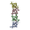
| ||||||||||||
|---|---|---|---|---|---|---|---|---|---|---|---|---|---|
| 1 |
| ||||||||||||
| Unit cell |
|
-Components
| #1: Protein | Mass: 34658.090 Da / Num. of mol.: 1 Source method: isolated from a genetically manipulated source Source: (gene. exp.) Gene: CDC10 / Plasmid: pACYC-Duet1 / Production host:  | ||
|---|---|---|---|
| #2: Protein | Mass: 38564.227 Da / Num. of mol.: 1 Source method: isolated from a genetically manipulated source Source: (gene. exp.) Gene: CDC3, YLR314C, L8543.7 / Plasmid: pACYC-Duet1 / Production host: | ||
| #3: Protein | Mass: 37554.496 Da / Num. of mol.: 1 Source method: isolated from a genetically manipulated source Source: (gene. exp.) Gene: CDC12 / Plasmid: pET-Duet1 / Production host: | ||
| #4: Protein | Mass: 36420.840 Da / Num. of mol.: 1 Source method: isolated from a genetically manipulated source Source: (gene. exp.) Gene: SHS1 / Plasmid: pET-Duet1 / Production host: | ||
| #5: Chemical | ChemComp-GDP /   Type: RNA linking / Mass: 443.201 Da / Num. of mol.: 4 / Source method: obtained synthetically / Formula: C10H15N5O11P2 / Feature type: SUBJECT OF INVESTIGATION / Comment: GDP, energy-carrying molecule*YM Type: RNA linking / Mass: 443.201 Da / Num. of mol.: 4 / Source method: obtained synthetically / Formula: C10H15N5O11P2 / Feature type: SUBJECT OF INVESTIGATION / Comment: GDP, energy-carrying molecule*YMHas ligand of interest | Y | |
-Experimental details
-Experiment
| Experiment | Method: X-RAY DIFFRACTION / Number of used crystals: 1 |
|---|
- Sample preparation
| Crystal | Density Matthews: 2.93 Å3/Da / Density % sol: 57.99 % |
|---|---|
| Crystal grow | Temperature: 293 K / Method: vapor diffusion, sitting drop / pH: 6.5 Details: 20 % PEG 5000, 0.3 M ammonium sulphate, 0.1 M BIS-TRIS pH 6.5 |
-Data collection
| Diffraction | Mean temperature: 100 K / Serial crystal experiment: N |
|---|---|
| Diffraction source | Source: SYNCHROTRON / Site: ESRF  / Beamline: ID30B / Wavelength: 0.885602 Å / Beamline: ID30B / Wavelength: 0.885602 Å |
| Detector | Type: DECTRIS EIGER X 9M / Detector: PIXEL / Date: Nov 6, 2022 |
| Radiation | Protocol: SINGLE WAVELENGTH / Monochromatic (M) / Laue (L): M / Scattering type: x-ray |
| Radiation wavelength | Wavelength: 0.885602 Å / Relative weight: 1 |
| Reflection | Resolution: 3.24→121.55 Å / Num. obs: 15695 / % possible obs: 63.2 % / Redundancy: 6.6 % / Biso Wilson estimate: 39.32 Å2 / CC1/2: 0.937 / Rpim(I) all: 0.334 / Net I/σ(I): 3.2 |
| Reflection shell | Resolution: 3.24→3.65 Å / Num. unique obs: 786 / CC1/2: 0.579 / Rpim(I) all: 0.877 / % possible all: 10.7 |
- Processing
| Software |
| |||||||||||||||||||||||||||||||||||||||||||||||||||||||||||||||||||||||||||||||||||||||||||||||||||||||||||||||||||||||||||||
|---|---|---|---|---|---|---|---|---|---|---|---|---|---|---|---|---|---|---|---|---|---|---|---|---|---|---|---|---|---|---|---|---|---|---|---|---|---|---|---|---|---|---|---|---|---|---|---|---|---|---|---|---|---|---|---|---|---|---|---|---|---|---|---|---|---|---|---|---|---|---|---|---|---|---|---|---|---|---|---|---|---|---|---|---|---|---|---|---|---|---|---|---|---|---|---|---|---|---|---|---|---|---|---|---|---|---|---|---|---|---|---|---|---|---|---|---|---|---|---|---|---|---|---|---|---|---|
| Refinement | Method to determine structure: MOLECULAR REPLACEMENT / Resolution: 3.24→121.55 Å / SU ML: 0.3423 / Cross valid method: FREE R-VALUE / σ(F): 1.34 / Phase error: 27.6657 Stereochemistry target values: GeoStd + Monomer Library + CDL v1.2
| |||||||||||||||||||||||||||||||||||||||||||||||||||||||||||||||||||||||||||||||||||||||||||||||||||||||||||||||||||||||||||||
| Solvent computation | Shrinkage radii: 0.9 Å / VDW probe radii: 1.1 Å / Solvent model: FLAT BULK SOLVENT MODEL | |||||||||||||||||||||||||||||||||||||||||||||||||||||||||||||||||||||||||||||||||||||||||||||||||||||||||||||||||||||||||||||
| Displacement parameters | Biso mean: 48.79 Å2 | |||||||||||||||||||||||||||||||||||||||||||||||||||||||||||||||||||||||||||||||||||||||||||||||||||||||||||||||||||||||||||||
| Refinement step | Cycle: LAST / Resolution: 3.24→121.55 Å
| |||||||||||||||||||||||||||||||||||||||||||||||||||||||||||||||||||||||||||||||||||||||||||||||||||||||||||||||||||||||||||||
| Refine LS restraints |
| |||||||||||||||||||||||||||||||||||||||||||||||||||||||||||||||||||||||||||||||||||||||||||||||||||||||||||||||||||||||||||||
| LS refinement shell |
| |||||||||||||||||||||||||||||||||||||||||||||||||||||||||||||||||||||||||||||||||||||||||||||||||||||||||||||||||||||||||||||
| Refinement TLS params. | Method: refined / Refine-ID: X-RAY DIFFRACTION
| |||||||||||||||||||||||||||||||||||||||||||||||||||||||||||||||||||||||||||||||||||||||||||||||||||||||||||||||||||||||||||||
| Refinement TLS group | Refine-ID: X-RAY DIFFRACTION
|
