 Movie
Movie Controller

[English] 日本語
 Yorodumi
- PDB-8pf4: Crystal structure of Trypanosoma brucei trypanothione reductase i... -
+ Open data

- Basic information
| Entry | Database: PDB / ID: 8pf4 | |||||||||
|---|---|---|---|---|---|---|---|---|---|---|
| Title | Crystal structure of Trypanosoma brucei trypanothione reductase in complex with 4-(((5-((4-fluorophenethyl)carbamoyl)furan-2-yl)methyl)(4-fluorophenyl)carbamoyl)-1-methyl-1-(3-phenylpropyl)piperazin-1-ium | |||||||||
 Components Components | Trypanothione reductase | |||||||||
 Keywords Keywords | OXIDOREDUCTASE / oxidoreductase activity / nucleotide binding / flavoenzyme / inhibitor binding | |||||||||
| Function / homology |  Function and homology information Function and homology informationtrypanothione-disulfide reductase / trypanothione-disulfide reductase (NADPH) activity / glutathione-disulfide reductase (NADPH) activity / glutathione metabolic process / cell redox homeostasis / flavin adenine dinucleotide binding / cellular response to oxidative stress / mitochondrion / cytosol Similarity search - Function | |||||||||
| Biological species |  | |||||||||
| Method |  X-RAY DIFFRACTION / SYNCHROTRON / MOLECULAR REPLACEMENT / Resolution: 1.84 Å X-RAY DIFFRACTION / SYNCHROTRON / MOLECULAR REPLACEMENT / Resolution: 1.84 Å | |||||||||
 Authors Authors | Exertier, C. / Ilari, A. / Fiorillo, A. / Antonelli, L. | |||||||||
| Funding support |  Italy, 1items Italy, 1items
| |||||||||
 Citation Citation | Journal: J.Med.Chem. / Year: 2024 Title: Fragment Merging, Growing, and Linking Identify New Trypanothione Reductase Inhibitors for Leishmaniasis. Authors: Exertier, C. / Salerno, A. / Antonelli, L. / Fiorillo, A. / Ocello, R. / Seghetti, F. / Caciolla, J. / Uliassi, E. / Masetti, M. / Fiorentino, E. / Orsini, S. / Di Muccio, T. / Ilari, A. / Bolognesi, M.L. | |||||||||
| History |
|
- Structure visualization
| Structure viewer | Molecule: MolmilJmol/JSmol |
|---|
- Downloads & links
-Download
| PDBx/mmCIF format | 8pf4.cif.gz | 427.1 KB | Display | PDBx/mmCIF format |
|---|---|---|---|---|
| PDB format | pdb8pf4.ent.gz | 346.7 KB | Display | PDB format |
| PDBx/mmJSON format | 8pf4.json.gz | Tree view | PDBx/mmJSON format | |
| Others |  Other downloads Other downloads |
-Validation report
| Summary document | 8pf4_validation.pdf.gz | 2.3 MB | Display | wwPDB validaton report |
|---|---|---|---|---|
| Full document | 8pf4_full_validation.pdf.gz | 2.4 MB | Display | |
| Data in XML | 8pf4_validation.xml.gz | 98 KB | Display | |
| Data in CIF | 8pf4_validation.cif.gz | 135.1 KB | Display | |
| Arichive directory | https://data.pdbj.org/pub/pdb/validation_reports/pf/8pf4ftp://data.pdbj.org/pub/pdb/validation_reports/pf/8pf4 | HTTPS FTP |
-Related structure data

-Links
 PDBj
PDBj


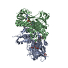
- Assembly
| Deposited unit | 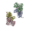
| ||||||||
|---|---|---|---|---|---|---|---|---|---|
| 1 | 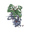
| ||||||||
| 2 | 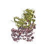
| ||||||||
| Unit cell |
|
-Components
-Protein , 1 types, 4 molecules ABCD
| #1: Protein | Mass: 53497.969 Da / Num. of mol.: 4 Source method: isolated from a genetically manipulated source Details: Owing to the flexibility of terminal segments, the electronic density is poorly defined which prevent us from being able to reconstruct the first G and the last four DSNL residues. Source: (gene. exp.)  References: UniProt: A0A3L6KZJ1, trypanothione-disulfide reductase |
|---|
-Non-polymers , 6 types, 1233 molecules 


| #2: Chemical | ChemComp-FAD /  Mass: 785.550 Da / Num. of mol.: 4 / Source method: obtained synthetically / Formula: C27H33N9O15P2 / Comment: FAD*YM Mass: 785.550 Da / Num. of mol.: 4 / Source method: obtained synthetically / Formula: C27H33N9O15P2 / Comment: FAD*YM#3: Chemical | ChemComp-YIA / ~{ Mass: 601.706 Da / Num. of mol.: 4 / Source method: obtained synthetically / Formula: C35H39F2N4O3 / Feature type: SUBJECT OF INVESTIGATION #4: Chemical | ChemComp-PEG /  Mass: 106.120 Da / Num. of mol.: 29 / Source method: obtained synthetically / Formula: C4H10O3 Mass: 106.120 Da / Num. of mol.: 29 / Source method: obtained synthetically / Formula: C4H10O3#5: Chemical | ChemComp-IMD /  Mass: 69.085 Da / Num. of mol.: 4 / Source method: obtained synthetically / Formula: C3H5N2 Mass: 69.085 Da / Num. of mol.: 4 / Source method: obtained synthetically / Formula: C3H5N2#6: Chemical | ChemComp-DMS /  Mass: 78.133 Da / Num. of mol.: 8 / Source method: obtained synthetically / Formula: C2H6OS / Comment: DMSO, precipitant*YM Mass: 78.133 Da / Num. of mol.: 8 / Source method: obtained synthetically / Formula: C2H6OS / Comment: DMSO, precipitant*YM#7: Water | ChemComp-HOH / | Mass: 18.015 Da / Num. of mol.: 1184 / Source method: isolated from a natural source / Formula: H2O |
|---|
-Details
| Has ligand of interest | Y |
|---|---|
| Has protein modification | Y |
-Experimental details
-Experiment
| Experiment | Method: X-RAY DIFFRACTION / Number of used crystals: 1 |
|---|
- Sample preparation
| Crystal | Density Matthews: 2.52 Å3/Da / Density % sol: 51.13 % |
|---|---|
| Crystal grow | Temperature: 293 K / Method: vapor diffusion, sitting drop Details: 13-15% PEG3350, 22-24% MPD, 40 mM imidazole pH 7.5, 50 mM NaBr |
-Data collection
| Diffraction | Mean temperature: 100 K / Serial crystal experiment: N |
|---|---|
| Diffraction source | Source: SYNCHROTRON / Site: ESRF  / Beamline: ID23-1 / Wavelength: 0.8856 Å / Beamline: ID23-1 / Wavelength: 0.8856 Å |
| Detector | Type: ADSC QUANTUM 315r / Detector: CCD / Date: Apr 5, 2023 |
| Radiation | Protocol: SINGLE WAVELENGTH / Monochromatic (M) / Laue (L): M / Scattering type: x-ray |
| Radiation wavelength | Wavelength: 0.8856 Å / Relative weight: 1 |
| Reflection | Resolution: 1.84→167.74 Å / Num. obs: 135585 / % possible obs: 94.7 % / Redundancy: 5.5 % / CC1/2: 0.994 / Net I/σ(I): 8.1 |
| Reflection shell | Resolution: 1.84→2.03 Å / Mean I/σ(I) obs: 1.6 / Num. unique obs: 6779 / CC1/2: 0.655 |
- Processing
| Software |
| ||||||||||||||||||||||||||||||||||||||||||||||||||||||||||||||||||||||||||||||||||||||||||||||||||||||||||||||||||||||||||||||||||||||||||||||||||||||||||||||||||||||||||||||||||||||
|---|---|---|---|---|---|---|---|---|---|---|---|---|---|---|---|---|---|---|---|---|---|---|---|---|---|---|---|---|---|---|---|---|---|---|---|---|---|---|---|---|---|---|---|---|---|---|---|---|---|---|---|---|---|---|---|---|---|---|---|---|---|---|---|---|---|---|---|---|---|---|---|---|---|---|---|---|---|---|---|---|---|---|---|---|---|---|---|---|---|---|---|---|---|---|---|---|---|---|---|---|---|---|---|---|---|---|---|---|---|---|---|---|---|---|---|---|---|---|---|---|---|---|---|---|---|---|---|---|---|---|---|---|---|---|---|---|---|---|---|---|---|---|---|---|---|---|---|---|---|---|---|---|---|---|---|---|---|---|---|---|---|---|---|---|---|---|---|---|---|---|---|---|---|---|---|---|---|---|---|---|---|---|---|
| Refinement | Method to determine structure: MOLECULAR REPLACEMENT / Resolution: 1.84→167.74 Å / Cor.coef. Fo:Fc: 0.951 / Cor.coef. Fo:Fc free: 0.918 / SU B: 4.276 / SU ML: 0.12 / Cross valid method: THROUGHOUT / ESU R: 0.197 / ESU R Free: 0.171 / Stereochemistry target values: MAXIMUM LIKELIHOOD / Details: HYDROGENS HAVE BEEN ADDED IN THE RIDING POSITIONS
| ||||||||||||||||||||||||||||||||||||||||||||||||||||||||||||||||||||||||||||||||||||||||||||||||||||||||||||||||||||||||||||||||||||||||||||||||||||||||||||||||||||||||||||||||||||||
| Solvent computation | Ion probe radii: 0.8 Å / Shrinkage radii: 0.8 Å / VDW probe radii: 1.2 Å / Solvent model: MASK | ||||||||||||||||||||||||||||||||||||||||||||||||||||||||||||||||||||||||||||||||||||||||||||||||||||||||||||||||||||||||||||||||||||||||||||||||||||||||||||||||||||||||||||||||||||||
| Displacement parameters | Biso mean: 22.457 Å2
| ||||||||||||||||||||||||||||||||||||||||||||||||||||||||||||||||||||||||||||||||||||||||||||||||||||||||||||||||||||||||||||||||||||||||||||||||||||||||||||||||||||||||||||||||||||||
| Refinement step | Cycle: 1 / Resolution: 1.84→167.74 Å
| ||||||||||||||||||||||||||||||||||||||||||||||||||||||||||||||||||||||||||||||||||||||||||||||||||||||||||||||||||||||||||||||||||||||||||||||||||||||||||||||||||||||||||||||||||||||
| Refine LS restraints |
|