+ Open data
- Basic information
| Entry | Database: PDB / ID: 8pcz | |||||||||||||||
|---|---|---|---|---|---|---|---|---|---|---|---|---|---|---|---|---|
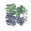
| Title | Ligand-free SpSLC9C1 in lipid nanodiscs, dimer | |||||||||||||||
 Components Components | Sperm-specific sodium proton exchanger | |||||||||||||||
 Keywords Keywords | MEMBRANE PROTEIN / SLC9 / NHE / sperm-specific | |||||||||||||||
| Function / homology |  Function and homology information Function and homology informationsperm head / potassium:proton antiporter activity / sodium:proton antiporter activity / sodium ion import across plasma membrane / cGMP binding / single fertilization / sperm flagellum / cAMP binding / potassium ion transmembrane transport / regulation of intracellular pH ...sperm head / potassium:proton antiporter activity / sodium:proton antiporter activity / sodium ion import across plasma membrane / cGMP binding / single fertilization / sperm flagellum / cAMP binding / potassium ion transmembrane transport / regulation of intracellular pH / protein homodimerization activity / plasma membrane Similarity search - Function | |||||||||||||||
| Biological species |  | |||||||||||||||
| Method | ELECTRON MICROSCOPY / single particle reconstruction / cryo EM / Resolution: 3.21 Å | |||||||||||||||
 Authors Authors | Kalienkova, V. / Peter, M. / Rheinberger, J. / Paulino, C. | |||||||||||||||
| Funding support |  Netherlands, Netherlands,  Switzerland, 4items Switzerland, 4items
| |||||||||||||||
 Citation Citation |  Journal: Nature / Year: 2023 Journal: Nature / Year: 2023Title: Structures of a sperm-specific solute carrier gated by voltage and cAMP. Authors: Valeria Kalienkova / Martin F Peter / Jan Rheinberger / Cristina Paulino /    Abstract: The newly characterized sperm-specific Na/H exchanger stands out by its unique tripartite domain composition. It unites a classical solute carrier unit with regulatory domains usually found in ion ...The newly characterized sperm-specific Na/H exchanger stands out by its unique tripartite domain composition. It unites a classical solute carrier unit with regulatory domains usually found in ion channels, namely, a voltage-sensing domain and a cyclic-nucleotide binding domain, which makes it a mechanistic chimera and a secondary-active transporter activated strictly by membrane voltage. Our structures of the sea urchin SpSLC9C1 in the absence and presence of ligands reveal the overall domain arrangement and new structural coupling elements. They allow us to propose a gating model, where movements in the voltage sensor indirectly cause the release of the exchanging unit from a locked state through long-distance allosteric effects transmitted by the newly characterized coupling helices. We further propose that modulation by its ligand cyclic AMP occurs by means of disruption of the cytosolic dimer interface, which lowers the energy barrier for S4 movements in the voltage-sensing domain. As SLC9C1 members have been shown to be essential for male fertility, including in mammals, our structure represents a potential new platform for the development of new on-demand contraceptives. | |||||||||||||||
| History |
|
- Structure visualization
| Structure viewer | Molecule:  Molmil Molmil Jmol/JSmol Jmol/JSmol |
|---|
- Downloads & links
- Download
| PDBx/mmCIF format |  8pcz.cif.gz 8pcz.cif.gz | 412.8 KB | Display |  PDBx/mmCIF format PDBx/mmCIF format |
|---|---|---|---|---|
| PDB format |  pdb8pcz.ent.gz pdb8pcz.ent.gz | 326.7 KB | Display |  PDB format PDB format |
| PDBx/mmJSON format |  8pcz.json.gz 8pcz.json.gz | Tree view |  PDBx/mmJSON format PDBx/mmJSON format | |
| Others |  Other downloads Other downloads |
-Validation report
| Arichive directory |  https://data.pdbj.org/pub/pdb/validation_reports/pc/8pcz https://data.pdbj.org/pub/pdb/validation_reports/pc/8pcz ftp://data.pdbj.org/pub/pdb/validation_reports/pc/8pcz ftp://data.pdbj.org/pub/pdb/validation_reports/pc/8pcz | HTTPS FTP |
|---|
-Related structure data
| Related structure data |  17596MC  8pd2C  8pd3C  8pd5C  8pd7C  8pd8C  8pd9C  8pduC  8pdvC M: map data used to model this data C: citing same article ( |
|---|---|
| Similar structure data | Similarity search - Function & homology  F&H Search F&H Search |
- Links
Links
- Assembly
| Deposited unit | 
|
|---|---|
| 1 |
|
- Components
| #1: Protein | Mass: 147624.484 Da / Num. of mol.: 2 Source method: isolated from a genetically manipulated source Source: (gene. exp.)  Cell line (production host): HEK293S GnTI- / Production host:  Homo sapiens (human) / References: UniProt: A3RL54 Homo sapiens (human) / References: UniProt: A3RL54 |
|---|
-Experimental details
-Experiment
| Experiment | Method: ELECTRON MICROSCOPY |
|---|---|
| EM experiment | Aggregation state: PARTICLE / 3D reconstruction method: single particle reconstruction |
- Sample preparation
| Component | Name: Ligand-free SpSLC9C1 in lipid nanodiscs, dimer / Type: COMPLEX / Entity ID: all / Source: RECOMBINANT | ||||||||||||
|---|---|---|---|---|---|---|---|---|---|---|---|---|---|
| Molecular weight | Value: 0.3 MDa / Experimental value: NO | ||||||||||||
| Source (natural) | Organism:  | ||||||||||||
| Source (recombinant) | Organism:  Homo sapiens (human) / Strain: HEK293S GnTI- Homo sapiens (human) / Strain: HEK293S GnTI- | ||||||||||||
| Buffer solution | pH: 7.6 | ||||||||||||
| Buffer component |
| ||||||||||||
| Specimen | Conc.: 0.93 mg/ml / Embedding applied: NO / Shadowing applied: NO / Staining applied: NO / Vitrification applied: YES | ||||||||||||
| Specimen support | Details: at 5 mA / Grid material: GOLD / Grid mesh size: 300 divisions/in. / Grid type: Quantifoil R1.2/1.3 | ||||||||||||
| Vitrification | Instrument: FEI VITROBOT MARK IV / Cryogen name: ETHANE-PROPANE / Humidity: 100 % / Chamber temperature: 288.15 K |
- Electron microscopy imaging
| Experimental equipment |  Model: Titan Krios / Image courtesy: FEI Company |
|---|---|
| Microscopy | Model: FEI TITAN KRIOS |
| Electron gun | Electron source:  FIELD EMISSION GUN / Accelerating voltage: 300 kV / Illumination mode: FLOOD BEAM FIELD EMISSION GUN / Accelerating voltage: 300 kV / Illumination mode: FLOOD BEAM |
| Electron lens | Mode: BRIGHT FIELD / Nominal magnification: 105000 X / Calibrated magnification: 59809 X / Nominal defocus max: 3000 nm / Nominal defocus min: 200 nm / Cs: 2.7 mm / C2 aperture diameter: 50 µm / Alignment procedure: COMA FREE |
| Specimen holder | Cryogen: NITROGEN / Specimen holder model: FEI TITAN KRIOS AUTOGRID HOLDER |
| Image recording | Average exposure time: 2.51 sec. / Electron dose: 60 e/Å2 / Film or detector model: GATAN K3 BIOQUANTUM (6k x 4k) / Num. of grids imaged: 1 / Num. of real images: 11299 |
| EM imaging optics | Energyfilter slit width: 20 eV |
- Processing
| EM software |
| ||||||||||||||||||||||||||||||||||||||||||||
|---|---|---|---|---|---|---|---|---|---|---|---|---|---|---|---|---|---|---|---|---|---|---|---|---|---|---|---|---|---|---|---|---|---|---|---|---|---|---|---|---|---|---|---|---|---|
| CTF correction | Type: PHASE FLIPPING AND AMPLITUDE CORRECTION | ||||||||||||||||||||||||||||||||||||||||||||
| Particle selection | Num. of particles selected: 5022107 | ||||||||||||||||||||||||||||||||||||||||||||
| Symmetry | Point symmetry: C2 (2 fold cyclic) | ||||||||||||||||||||||||||||||||||||||||||||
| 3D reconstruction | Resolution: 3.21 Å / Resolution method: FSC 0.143 CUT-OFF / Num. of particles: 1458809 / Num. of class averages: 1 / Symmetry type: POINT | ||||||||||||||||||||||||||||||||||||||||||||
| Refinement | Cross valid method: NONE | ||||||||||||||||||||||||||||||||||||||||||||
| Refine LS restraints |
|
 Movie
Movie Controller


 PDBj
PDBj


