 Movie
Movie Controller

[English] 日本語
 Yorodumi
- PDB-8paq: Structure of the small subunit of the laccase-like Nlac protein f... -
+ Open data

- Basic information
| Entry | Database: PDB / ID: 8paq | |||||||||
|---|---|---|---|---|---|---|---|---|---|---|
| Title | Structure of the small subunit of the laccase-like Nlac protein from Pleurotus eryngii | |||||||||
 Components Components | POXA3b laccase small subunit | |||||||||
 Keywords Keywords | OXIDOREDUCTASE / NLac / PeNL / Pleurotus eryngii / Small subunit / Laccase-like enzyme | |||||||||
| Function / homology | hydroquinone:oxygen oxidoreductase activity / laccase / IMIDAZOLE / POXA3b laccase small subunit Function and homology information Function and homology information | |||||||||
| Biological species |  Pleurotus eryngii (fungus) Pleurotus eryngii (fungus) | |||||||||
| Method |  X-RAY DIFFRACTION / SYNCHROTRON / MOLECULAR REPLACEMENT / Resolution: 1.6 Å X-RAY DIFFRACTION / SYNCHROTRON / MOLECULAR REPLACEMENT / Resolution: 1.6 Å | |||||||||
 Authors Authors | Medrano, F.J. / Camarero, S. | |||||||||
| Funding support |  Spain, 2items Spain, 2items
| |||||||||
 Citation Citation | Journal: Protein Sci. / Year: 2023 Title: Role and structure of the small subunit forming heterodimers with laccase-like enzymes. Authors: Aza, P. / Linde, D. / Molpeceres, G. / Vind, J. / Medrano, F.J. / Camarero, S. | |||||||||
| History |
|
- Structure visualization
| Structure viewer | Molecule: MolmilJmol/JSmol |
|---|
- Downloads & links
-Download
| PDBx/mmCIF format | 8paq.cif.gz | 94.6 KB | Display | PDBx/mmCIF format |
|---|---|---|---|---|
| PDB format | pdb8paq.ent.gz | 58.4 KB | Display | PDB format |
| PDBx/mmJSON format | 8paq.json.gz | Tree view | PDBx/mmJSON format | |
| Others |  Other downloads Other downloads |
-Validation report
| Summary document | 8paq_validation.pdf.gz | 443 KB | Display | wwPDB validaton report |
|---|---|---|---|---|
| Full document | 8paq_full_validation.pdf.gz | 443.3 KB | Display | |
| Data in XML | 8paq_validation.xml.gz | 16.4 KB | Display | |
| Data in CIF | 8paq_validation.cif.gz | 24 KB | Display | |
| Arichive directory | https://data.pdbj.org/pub/pdb/validation_reports/pa/8paqftp://data.pdbj.org/pub/pdb/validation_reports/pa/8paq | HTTPS FTP |
-Related structure data
| Similar structure data |
|---|
-Links
 PDBj
PDBj


- Assembly
| Deposited unit | 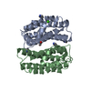
| ||||||||||||
|---|---|---|---|---|---|---|---|---|---|---|---|---|---|
| 1 | 
| ||||||||||||
| 2 | 
| ||||||||||||
| Unit cell |
|
-Components
| #1: Protein | Mass: 17425.416 Da / Num. of mol.: 2 Source method: isolated from a genetically manipulated source Source: (gene. exp.) Pleurotus eryngii (fungus) / Production host:  #2: Chemical | ChemComp-CA / |   Mass: 40.078 Da / Num. of mol.: 1 / Source method: obtained synthetically / Formula: Ca Mass: 40.078 Da / Num. of mol.: 1 / Source method: obtained synthetically / Formula: Ca#3: Chemical | ChemComp-IMD /   Mass: 69.085 Da / Num. of mol.: 4 / Source method: obtained synthetically / Formula: C3H5N2 Mass: 69.085 Da / Num. of mol.: 4 / Source method: obtained synthetically / Formula: C3H5N2#4: Chemical | ChemComp-EDO / |   Mass: 62.068 Da / Num. of mol.: 1 / Source method: obtained synthetically / Formula: C2H6O2 Mass: 62.068 Da / Num. of mol.: 1 / Source method: obtained synthetically / Formula: C2H6O2#5: Water | ChemComp-HOH / |  Mass: 18.015 Da / Num. of mol.: 315 / Source method: isolated from a natural source / Formula: H2O Mass: 18.015 Da / Num. of mol.: 315 / Source method: isolated from a natural source / Formula: H2OHas ligand of interest | N | |
|---|
-Experimental details
-Experiment
| Experiment | Method: X-RAY DIFFRACTION / Number of used crystals: 1 |
|---|
- Sample preparation
| Crystal | Density Matthews: 3.11 Å3/Da / Density % sol: 60.5 % |
|---|---|
| Crystal grow | Temperature: 295 K / Method: vapor diffusion, sitting drop / pH: 8 / Details: 0.1 M Imidazole at pH 8.0 and 2.5 M NaCl |
-Data collection
| Diffraction | Mean temperature: 100 K / Serial crystal experiment: N |
|---|---|
| Diffraction source | Source: SYNCHROTRON / Site: ALBA / Beamline: XALOC / Wavelength: 0.979257 Å |
| Detector | Type: DECTRIS PILATUS 6M-F / Detector: PIXEL / Date: Nov 24, 2022 |
| Radiation | Protocol: SINGLE WAVELENGTH / Monochromatic (M) / Laue (L): M / Scattering type: x-ray |
| Radiation wavelength | Wavelength: 0.979257 Å / Relative weight: 1 |
| Reflection | Resolution: 1.6→47.16 Å / Num. obs: 58835 / % possible obs: 99.9 % / Redundancy: 26.2 % / Biso Wilson estimate: 25.78 Å2 / CC1/2: 0.999 / Net I/σ(I): 14.29 |
| Reflection shell | Resolution: 1.6→1.69 Å / Mean I/σ(I) obs: 0.69 / Num. unique obs: 9324 / CC1/2: 0.385 / % possible all: 99.3 |
- Processing
| Software |
| |||||||||||||||||||||||||||||||||||||||||||||||||||||||||||||||||||||||||||||||||||||||||||||||||||||||||
|---|---|---|---|---|---|---|---|---|---|---|---|---|---|---|---|---|---|---|---|---|---|---|---|---|---|---|---|---|---|---|---|---|---|---|---|---|---|---|---|---|---|---|---|---|---|---|---|---|---|---|---|---|---|---|---|---|---|---|---|---|---|---|---|---|---|---|---|---|---|---|---|---|---|---|---|---|---|---|---|---|---|---|---|---|---|---|---|---|---|---|---|---|---|---|---|---|---|---|---|---|---|---|---|---|---|---|
| Refinement | Method to determine structure: MOLECULAR REPLACEMENT / Resolution: 1.6→47.16 Å / SU ML: 0.2378 / Cross valid method: FREE R-VALUE / σ(F): 1.33 / Phase error: 24.2677 Stereochemistry target values: GeoStd + Monomer Library + CDL v1.2
| |||||||||||||||||||||||||||||||||||||||||||||||||||||||||||||||||||||||||||||||||||||||||||||||||||||||||
| Solvent computation | Shrinkage radii: 0.9 Å / VDW probe radii: 1.1 Å / Solvent model: FLAT BULK SOLVENT MODEL | |||||||||||||||||||||||||||||||||||||||||||||||||||||||||||||||||||||||||||||||||||||||||||||||||||||||||
| Displacement parameters | Biso mean: 30.88 Å2 | |||||||||||||||||||||||||||||||||||||||||||||||||||||||||||||||||||||||||||||||||||||||||||||||||||||||||
| Refinement step | Cycle: LAST / Resolution: 1.6→47.16 Å
| |||||||||||||||||||||||||||||||||||||||||||||||||||||||||||||||||||||||||||||||||||||||||||||||||||||||||
| Refine LS restraints |
| |||||||||||||||||||||||||||||||||||||||||||||||||||||||||||||||||||||||||||||||||||||||||||||||||||||||||
| LS refinement shell |
|
