 Movie
Movie Controller

[English] 日本語
 Yorodumi
- PDB-8p8x: Crystal structure of a pathogenic mutant variant of human mitocho... -
+ Open data

- Basic information
| Entry | Database: PDB / ID: 8p8x | ||||||
|---|---|---|---|---|---|---|---|
| Title | Crystal structure of a pathogenic mutant variant of human mitochodnrial PheRS | ||||||
 Components Components | Phenylalanine--tRNA ligase, mitochondrial | ||||||
 Keywords Keywords | LIGASE / Phenylalanyl-tRNA synthetase / FARS2 / mitochondria / mitochondrial disease / class II aminoacyl-tRNA ligase / alpha-beta domain / ATP binding / amino acid binding | ||||||
| Function / homology |  Function and homology information Function and homology informationphenylalanine-tRNA ligase / Mitochondrial tRNA aminoacylation / phenylalanyl-tRNA aminoacylation / phenylalanine-tRNA ligase activity / tRNA aminoacylation for protein translation / tRNA processing / tRNA binding / mitochondrial matrix / mitochondrion / ATP binding / cytoplasm Similarity search - Function | ||||||
| Biological species |  Homo sapiens (human) Homo sapiens (human) | ||||||
| Method |  X-RAY DIFFRACTION / SYNCHROTRON / MOLECULAR REPLACEMENT / Resolution: 1.46 Å X-RAY DIFFRACTION / SYNCHROTRON / MOLECULAR REPLACEMENT / Resolution: 1.46 Å | ||||||
 Authors Authors | Chen, W. / Kuhle, B. | ||||||
| Funding support | 1items
| ||||||
 Citation Citation | Journal: Mol.Genet.Metab. / Year: 2023 Title: Clinical and molecular characterization of novel FARS2 variants causing neonatal mitochondrial disease. Authors: Chen, W. / Rehsi, P. / Thompson, K. / Yeo, M. / Stals, K. / He, L. / Schimmel, P. / Chrzanowska-Lightowlers, Z.M.A. / Wakeling, E. / Taylor, R.W. / Kuhle, B. | ||||||
| History |
|
- Structure visualization
| Structure viewer | Molecule: MolmilJmol/JSmol |
|---|
- Downloads & links
-Download
| PDBx/mmCIF format | 8p8x.cif.gz | 191.4 KB | Display | PDBx/mmCIF format |
|---|---|---|---|---|
| PDB format | pdb8p8x.ent.gz | 149 KB | Display | PDB format |
| PDBx/mmJSON format | 8p8x.json.gz | Tree view | PDBx/mmJSON format | |
| Others |  Other downloads Other downloads |
-Validation report
| Arichive directory | https://data.pdbj.org/pub/pdb/validation_reports/p8/8p8xftp://data.pdbj.org/pub/pdb/validation_reports/p8/8p8x | HTTPS FTP |
|---|
-Related structure data
| Similar structure data |
|---|
-Links
 PDBj
PDBj


- Assembly
| Deposited unit | 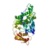
| ||||||||
|---|---|---|---|---|---|---|---|---|---|
| 1 |
| ||||||||
| Unit cell |
|
-Components
| #1: Protein | Mass: 48413.902 Da / Num. of mol.: 1 Source method: isolated from a genetically manipulated source Source: (gene. exp.) Homo sapiens (human) / Gene: FARS2, FARS1, HSPC320 / Production host:  |
|---|---|
| #2: Chemical | ChemComp-PHE /   Type: L-peptide linking / Mass: 165.189 Da / Num. of mol.: 1 / Source method: obtained synthetically / Formula: C9H11NO2 Type: L-peptide linking / Mass: 165.189 Da / Num. of mol.: 1 / Source method: obtained synthetically / Formula: C9H11NO2 |
| #3: Water | ChemComp-HOH /  Mass: 18.015 Da / Num. of mol.: 477 / Source method: isolated from a natural source / Formula: H2O Mass: 18.015 Da / Num. of mol.: 477 / Source method: isolated from a natural source / Formula: H2O |
| Has ligand of interest | N |
-Experimental details
-Experiment
| Experiment | Method: X-RAY DIFFRACTION / Number of used crystals: 1 |
|---|
- Sample preparation
| Crystal | Density Matthews: 2.43 Å3/Da / Density % sol: 49.29 % |
|---|---|
| Crystal grow | Temperature: 296 K / Method: vapor diffusion, sitting drop / pH: 7 Details: 1.7 M sodium acetate trihydrate pH 7, 0.1 M BIS-TRIS propane pH 7, 0.1 M MgCl2, 1% v/v 1,2-Butanediol |
-Data collection
| Diffraction | Mean temperature: 100 K / Serial crystal experiment: N |
|---|---|
| Diffraction source | Source: SYNCHROTRON / Site: SSRL  / Beamline: BL12-1 / Wavelength: 0.9795 Å / Beamline: BL12-1 / Wavelength: 0.9795 Å |
| Detector | Type: DECTRIS EIGER X 16M / Detector: PIXEL / Date: Mar 25, 2022 |
| Radiation | Protocol: SINGLE WAVELENGTH / Monochromatic (M) / Laue (L): M / Scattering type: x-ray |
| Radiation wavelength | Wavelength: 0.9795 Å / Relative weight: 1 |
| Reflection | Resolution: 1.46→36.32 Å / Num. obs: 82262 / % possible obs: 99.7 % / Redundancy: 6.6 % / CC1/2: 1 / Rmerge(I) obs: 0.039 / Rpim(I) all: 0.016 / Rrim(I) all: 0.042 / Χ2: 0.95 / Net I/σ(I): 21 / Num. measured all: 546378 |
| Reflection shell | Resolution: 1.46→1.48 Å / % possible obs: 95.3 % / Redundancy: 4.6 % / Rmerge(I) obs: 0.631 / Num. measured all: 17715 / Num. unique obs: 3860 / CC1/2: 0.792 / Rpim(I) all: 0.323 / Rrim(I) all: 0.713 / Χ2: 0.9 / Net I/σ(I) obs: 2 |
- Processing
| Software |
| |||||||||||||||||||||||||||||||||||||||||||||||||||||||||||||||||||||||||||||||||||||||||||||||||||||||||
|---|---|---|---|---|---|---|---|---|---|---|---|---|---|---|---|---|---|---|---|---|---|---|---|---|---|---|---|---|---|---|---|---|---|---|---|---|---|---|---|---|---|---|---|---|---|---|---|---|---|---|---|---|---|---|---|---|---|---|---|---|---|---|---|---|---|---|---|---|---|---|---|---|---|---|---|---|---|---|---|---|---|---|---|---|---|---|---|---|---|---|---|---|---|---|---|---|---|---|---|---|---|---|---|---|---|---|
| Refinement | Method to determine structure: MOLECULAR REPLACEMENT / Resolution: 1.46→36.32 Å / SU ML: 0.14 / Cross valid method: FREE R-VALUE / σ(F): 1.34 / Phase error: 16.63 / Stereochemistry target values: ML
| |||||||||||||||||||||||||||||||||||||||||||||||||||||||||||||||||||||||||||||||||||||||||||||||||||||||||
| Solvent computation | Shrinkage radii: 0.9 Å / VDW probe radii: 1.11 Å / Solvent model: FLAT BULK SOLVENT MODEL | |||||||||||||||||||||||||||||||||||||||||||||||||||||||||||||||||||||||||||||||||||||||||||||||||||||||||
| Refinement step | Cycle: LAST / Resolution: 1.46→36.32 Å
| |||||||||||||||||||||||||||||||||||||||||||||||||||||||||||||||||||||||||||||||||||||||||||||||||||||||||
| Refine LS restraints |
| |||||||||||||||||||||||||||||||||||||||||||||||||||||||||||||||||||||||||||||||||||||||||||||||||||||||||
| LS refinement shell |
|
