 Movie
Movie Controller


+ Open data

- Basic information
| Entry | Database: PDB / ID: 8p8q | |||||||||
|---|---|---|---|---|---|---|---|---|---|---|
| Title | Recombinant Ym1 crystal structure | |||||||||
 Components Components | Chitinase-like protein 3 | |||||||||
 Keywords Keywords | IMMUNE SYSTEM / Ym1 / Chil3 / Chitinase-like protein | |||||||||
| Function / homology |  Function and homology information Function and homology informationrough endoplasmic reticulum lumen / beta-N-acetylhexosaminidase activity / beta-N-acetylhexosaminidase / response to selenium ion / response to nematode / chitin binding / polysaccharide catabolic process / nuclear envelope / carbohydrate binding / cytoplasmic vesicle ...rough endoplasmic reticulum lumen / beta-N-acetylhexosaminidase activity / beta-N-acetylhexosaminidase / response to selenium ion / response to nematode / chitin binding / polysaccharide catabolic process / nuclear envelope / carbohydrate binding / cytoplasmic vesicle / inflammatory response / extracellular region / cytoplasm Similarity search - Function | |||||||||
| Biological species |  | |||||||||
| Method |  X-RAY DIFFRACTION / SYNCHROTRON / MOLECULAR REPLACEMENT / Resolution: 1.792 Å X-RAY DIFFRACTION / SYNCHROTRON / MOLECULAR REPLACEMENT / Resolution: 1.792 Å | |||||||||
 Authors Authors | Verschueren, K.H.G. / Verstraete, K. / Heyndrickx, I. / Aegerter, H. / Smole, U. / Savvides, S.N. / Lambrecht, B.N. | |||||||||
| Funding support | European Union,  Belgium, 2items Belgium, 2items
| |||||||||
 Citation Citation | Journal: Elife / Year: 2024 Title: Ym1 protein crystals promote type 2 immunity. Authors: Heyndrickx, I. / Deswarte, K. / Verstraete, K. / Verschueren, K.H.G. / Smole, U. / Aegerter, H. / Dansercoer, A. / Hammad, H. / Savvides, S.N. / Lambrecht, B.N. #1: Journal: Elife / Year: 2023Title: Ym1 protein crystals promote type 2 immunity Authors: Heyndrickx, I. / Deswarte, K. / Verstraete, K. / Verschueren, K. / Smole, U. / Aegerter, H. / Dansercoer, A. / Hammad, H. / Savvides, S. / Lambrecht, B. | |||||||||
| History |
|
- Structure visualization
| Structure viewer | Molecule: MolmilJmol/JSmol |
|---|
- Downloads & links
-Download
| PDBx/mmCIF format | 8p8q.cif.gz | 165.1 KB | Display | PDBx/mmCIF format |
|---|---|---|---|---|
| PDB format | pdb8p8q.ent.gz | 127.4 KB | Display | PDB format |
| PDBx/mmJSON format | 8p8q.json.gz | Tree view | PDBx/mmJSON format | |
| Others |  Other downloads Other downloads |
-Validation report
| Arichive directory | https://data.pdbj.org/pub/pdb/validation_reports/p8/8p8qftp://data.pdbj.org/pub/pdb/validation_reports/p8/8p8q | HTTPS FTP |
|---|
-Related structure data


-Links
 PDBj
PDBj- Assembly
| Deposited unit | 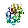
| ||||||||
|---|---|---|---|---|---|---|---|---|---|
| 1 |
| ||||||||
| Unit cell |
|
-Components
| #1: Protein | Mass: 44388.898 Da / Num. of mol.: 1 Source method: isolated from a genetically manipulated source Details: Recombinant Ym1 (Uniprot ID O35744, residues 22 - 398) was secreted from HEK293 FreeStyle cells and purified via ion exchange chromatography and SEC. The mouse IgH signal peptide ...Details: Recombinant Ym1 (Uniprot ID O35744, residues 22 - 398) was secreted from HEK293 FreeStyle cells and purified via ion exchange chromatography and SEC. The mouse IgH signal peptide (MGWSCIIFFLVATATGVHS) was used as a secretion signal. Source: (gene. exp.) Details (production host): Ym1 residues 22 to 398 in frame with the mouse IgH signal peptide Cell line (production host): HEK293 FreeStyle / Production host:  Homo sapiens (human) / References: UniProt: O35744, beta-N-acetylhexosaminidase Homo sapiens (human) / References: UniProt: O35744, beta-N-acetylhexosaminidase |
|---|---|
| #2: Chemical | ChemComp-ACT /   Mass: 59.044 Da / Num. of mol.: 1 / Source method: obtained synthetically / Formula: C2H3O2 Mass: 59.044 Da / Num. of mol.: 1 / Source method: obtained synthetically / Formula: C2H3O2 |
| #3: Chemical | ChemComp-GOL /   Mass: 92.094 Da / Num. of mol.: 1 / Source method: obtained synthetically / Formula: C3H8O3 Mass: 92.094 Da / Num. of mol.: 1 / Source method: obtained synthetically / Formula: C3H8O3 |
| #4: Water | ChemComp-HOH /  Mass: 18.015 Da / Num. of mol.: 220 / Source method: isolated from a natural source / Formula: H2O Mass: 18.015 Da / Num. of mol.: 220 / Source method: isolated from a natural source / Formula: H2O |
| Has ligand of interest | N |
| Has protein modification | Y |
-Experimental details
-Experiment
| Experiment | Method: X-RAY DIFFRACTION / Number of used crystals: 1 |
|---|
- Sample preparation
| Crystal | Density Matthews: 2.04 Å3/Da / Density % sol: 39.66 % |
|---|---|
| Crystal grow | Temperature: 293 K / Method: vapor diffusion, sitting drop / pH: 6.5 Details: Condition A7 of the Hampton Research Crystal Screen HT (0.1 M sodium cacodylate pH 6.5, 1.4 M sodium acetate) Cryo-protection: mother liquor supplemented with 30% (v/v) glycerol |
-Data collection
| Diffraction | Mean temperature: 100 K / Ambient temp details: Cold N2 gas stream / Serial crystal experiment: N |
|---|---|
| Diffraction source | Source: SYNCHROTRON / Site: SOLEIL  / Beamline: PROXIMA 1 / Wavelength: 0.97857 Å / Beamline: PROXIMA 1 / Wavelength: 0.97857 Å |
| Detector | Type: DECTRIS PILATUS 6M / Detector: PIXEL / Date: Jul 5, 2018 |
| Radiation | Protocol: SINGLE WAVELENGTH / Monochromatic (M) / Laue (L): M / Scattering type: x-ray |
| Radiation wavelength | Wavelength: 0.97857 Å / Relative weight: 1 |
| Reflection | Resolution: 1.79→59.92 Å / Num. obs: 33407 / % possible obs: 99.2 % / Redundancy: 4.06 % / Biso Wilson estimate: 20.318 Å2 / CC1/2: 0.979 / Rrim(I) all: 0.247 / Net I/σ(I): 5.28 |
| Reflection shell | Resolution: 1.79→1.9 Å / Mean I/σ(I) obs: 1.54 / Num. unique obs: 5172 / CC1/2: 0.566 / Rrim(I) all: 1.121 / % possible all: 96 |
- Processing
| Software |
| ||||||||||||||||||||||||||||||||||||||||||||||||||||||||||||||||||||||||||||||||||||||||||||||||||||||||||||||||||||||||||||||||||||||||||||||||||||||||||||||||||||||||||||||||||||||||||||||||||||||||
|---|---|---|---|---|---|---|---|---|---|---|---|---|---|---|---|---|---|---|---|---|---|---|---|---|---|---|---|---|---|---|---|---|---|---|---|---|---|---|---|---|---|---|---|---|---|---|---|---|---|---|---|---|---|---|---|---|---|---|---|---|---|---|---|---|---|---|---|---|---|---|---|---|---|---|---|---|---|---|---|---|---|---|---|---|---|---|---|---|---|---|---|---|---|---|---|---|---|---|---|---|---|---|---|---|---|---|---|---|---|---|---|---|---|---|---|---|---|---|---|---|---|---|---|---|---|---|---|---|---|---|---|---|---|---|---|---|---|---|---|---|---|---|---|---|---|---|---|---|---|---|---|---|---|---|---|---|---|---|---|---|---|---|---|---|---|---|---|---|---|---|---|---|---|---|---|---|---|---|---|---|---|---|---|---|---|---|---|---|---|---|---|---|---|---|---|---|---|---|---|---|---|
| Refinement | Method to determine structure: MOLECULAR REPLACEMENT / Resolution: 1.792→42.4 Å / Cor.coef. Fo:Fc: 0.921 / Cor.coef. Fo:Fc free: 0.894 / SU R Cruickshank DPI: 0.122 / Cross valid method: THROUGHOUT / σ(F): 0 / SU R Blow DPI: 0.124 / SU Rfree Blow DPI: 0.11 / SU Rfree Cruickshank DPI: 0.11
| ||||||||||||||||||||||||||||||||||||||||||||||||||||||||||||||||||||||||||||||||||||||||||||||||||||||||||||||||||||||||||||||||||||||||||||||||||||||||||||||||||||||||||||||||||||||||||||||||||||||||
| Displacement parameters | Biso mean: 12.8 Å2
| ||||||||||||||||||||||||||||||||||||||||||||||||||||||||||||||||||||||||||||||||||||||||||||||||||||||||||||||||||||||||||||||||||||||||||||||||||||||||||||||||||||||||||||||||||||||||||||||||||||||||
| Refine analyze | Luzzati coordinate error obs: 0.23 Å | ||||||||||||||||||||||||||||||||||||||||||||||||||||||||||||||||||||||||||||||||||||||||||||||||||||||||||||||||||||||||||||||||||||||||||||||||||||||||||||||||||||||||||||||||||||||||||||||||||||||||
| Refinement step | Cycle: LAST / Resolution: 1.792→42.4 Å
| ||||||||||||||||||||||||||||||||||||||||||||||||||||||||||||||||||||||||||||||||||||||||||||||||||||||||||||||||||||||||||||||||||||||||||||||||||||||||||||||||||||||||||||||||||||||||||||||||||||||||
| Refine LS restraints |
| ||||||||||||||||||||||||||||||||||||||||||||||||||||||||||||||||||||||||||||||||||||||||||||||||||||||||||||||||||||||||||||||||||||||||||||||||||||||||||||||||||||||||||||||||||||||||||||||||||||||||
| LS refinement shell | Resolution: 1.792→1.81 Å
| ||||||||||||||||||||||||||||||||||||||||||||||||||||||||||||||||||||||||||||||||||||||||||||||||||||||||||||||||||||||||||||||||||||||||||||||||||||||||||||||||||||||||||||||||||||||||||||||||||||||||
| Refinement TLS params. | Method: refined / Refine-ID: X-RAY DIFFRACTION
| ||||||||||||||||||||||||||||||||||||||||||||||||||||||||||||||||||||||||||||||||||||||||||||||||||||||||||||||||||||||||||||||||||||||||||||||||||||||||||||||||||||||||||||||||||||||||||||||||||||||||
| Refinement TLS group |
|
