Deposited unit
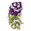
A: Twin-arginine translocation signal domain-containing protein
D: Twin-arginine translocation signal domain-containing protein
G: Twin-arginine translocation signal domain-containing protein
J: Twin-arginine translocation signal domain-containing protein
M: Twin-arginine translocation signal domain-containing protein
P: Twin-arginine translocation signal domain-containing protein
S: Twin-arginine translocation signal domain-containing protein
V: Twin-arginine translocation signal domain-containing protein
Y: Twin-arginine translocation signal domain-containing protein
2: Twin-arginine translocation signal domain-containing protein
5: Twin-arginine translocation signal domain-containing protein
8: Twin-arginine translocation signal domain-containing protein
x: Twin-arginine translocation signal domain-containing protein
e: Twin-arginine translocation signal domain-containing protein
h: Twin-arginine translocation signal domain-containing protein
k: Twin-arginine translocation signal domain-containing protein
hetero molecules Summary Component details
Theoretical mass Number of molelcules Total (without water) 882,950 69 Polymers 879,114 16 Non-polymers 3,836 53 Water 43,596 2420
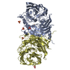
1
A: Twin-arginine translocation signal domain-containing protein
D: Twin-arginine translocation signal domain-containing protein
hetero molecules Summary Component details Symmetry operations Calculated values
Theoretical mass Number of molelcules Total (without water) 110,555 11 Polymers 109,889 2 Non-polymers 666 9 Water 36 2
Type Name Symmetry operation Number identity operation 1_555 x,y,z 1
Buried area 6410 Å2 ΔGint -116 kcal/mol Surface area 28520 Å2 Method
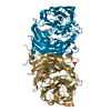
2
G: Twin-arginine translocation signal domain-containing protein
J: Twin-arginine translocation signal domain-containing protein
hetero molecules Summary Component details Symmetry operations Calculated values
Theoretical mass Number of molelcules Total (without water) 110,335 8 Polymers 109,889 2 Non-polymers 446 6 Water 36 2
Type Name Symmetry operation Number identity operation 1_555 x,y,z 1
Buried area 6050 Å2 ΔGint -109 kcal/mol Surface area 28340 Å2 Method
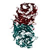
3
M: Twin-arginine translocation signal domain-containing protein
P: Twin-arginine translocation signal domain-containing protein
hetero molecules Summary Component details Symmetry operations Calculated values
Theoretical mass Number of molelcules Total (without water) 110,455 10 Polymers 109,889 2 Non-polymers 565 8 Water 36 2
Type Name Symmetry operation Number identity operation 1_555 x,y,z 1
Buried area 6200 Å2 ΔGint -118 kcal/mol Surface area 28280 Å2 Method
4
S: Twin-arginine translocation signal domain-containing protein
V: Twin-arginine translocation signal domain-containing protein
hetero molecules Summary Component details Symmetry operations Calculated values
Theoretical mass Number of molelcules Total (without water) 110,358 9 Polymers 109,889 2 Non-polymers 469 7 Water 36 2
Type Name Symmetry operation Number identity operation 1_555 x,y,z 1
Buried area 6070 Å2 ΔGint -108 kcal/mol Surface area 28050 Å2 Method
5
Y: Twin-arginine translocation signal domain-containing protein
2: Twin-arginine translocation signal domain-containing protein
hetero molecules Summary Component details Symmetry operations Calculated values
Theoretical mass Number of molelcules Total (without water) 110,335 8 Polymers 109,889 2 Non-polymers 446 6 Water 36 2
Type Name Symmetry operation Number identity operation 1_555 x,y,z 1
Buried area 6010 Å2 ΔGint -107 kcal/mol Surface area 28260 Å2 Method
6
5: Twin-arginine translocation signal domain-containing protein
8: Twin-arginine translocation signal domain-containing protein
hetero molecules Summary Component details Symmetry operations Calculated values
Theoretical mass Number of molelcules Total (without water) 110,335 8 Polymers 109,889 2 Non-polymers 446 6 Water 36 2
Type Name Symmetry operation Number identity operation 1_555 x,y,z 1
Buried area 6070 Å2 ΔGint -107 kcal/mol Surface area 28200 Å2 Method
7
x: Twin-arginine translocation signal domain-containing protein
e: Twin-arginine translocation signal domain-containing protein
hetero molecules Summary Component details Symmetry operations Calculated values
Theoretical mass Number of molelcules Total (without water) 110,239 7 Polymers 109,889 2 Non-polymers 350 5 Water 36 2
Type Name Symmetry operation Number identity operation 1_555 x,y,z 1
Buried area 5870 Å2 ΔGint -95 kcal/mol Surface area 28210 Å2 Method
8
h: Twin-arginine translocation signal domain-containing protein
k: Twin-arginine translocation signal domain-containing protein
hetero molecules Summary Component details Symmetry operations Calculated values
Theoretical mass Number of molelcules Total (without water) 110,335 8 Polymers 109,889 2 Non-polymers 446 6 Water 36 2
Type Name Symmetry operation Number identity operation 1_555 x,y,z 1
Buried area 6030 Å2 ΔGint -105 kcal/mol Surface area 28190 Å2 Method
Unit cell Length a, b, c (Å) 98.150, 142.420, 294.400 Angle α, β, γ (deg.) 90.00, 90.07, 90.00 Int Tables number 4 Space group name H-M P121 1
 Movie
Movie Controller

 Yorodumi
 Open data
 Basic information
 Components
 Keywords
 Function and homology information
 Thioalkalivibrio paradoxus ARh 1 (bacteria)
 X-RAY DIFFRACTION /
  Authors
 Russian Federation, 1items
  Citation
 Structure visualization
 Downloads & links
 Other downloads
 PDBj
PDBj

 Assembly


 Mass: 96.063 Da / Num. of mol.: 17 / Source method: obtained synthetically / Formula: SO4
 Mass: 61.833 Da / Num. of mol.: 2 / Source method: obtained synthetically / Formula: BH3O3
 Mass: 63.546 Da / Num. of mol.: 32 / Source method: obtained synthetically / Formula: Cu / Feature type: SUBJECT OF INVESTIGATION
 Mass: 22.990 Da / Num. of mol.: 2 / Source method: obtained synthetically / Formula: Na
 Sample preparation
 / Beamline: BL41XU / Wavelength: 1 Å
 Processing