 Movie
Movie Controller


+ Open data

- Basic information
| Entry | Database: PDB / ID: 8p1u | |||||||||||||||||||||||||||||||||||||||
|---|---|---|---|---|---|---|---|---|---|---|---|---|---|---|---|---|---|---|---|---|---|---|---|---|---|---|---|---|---|---|---|---|---|---|---|---|---|---|---|---|
| Title | Structure of divisome complex FtsWIQLB | |||||||||||||||||||||||||||||||||||||||
 Components Components |
| |||||||||||||||||||||||||||||||||||||||
 Keywords Keywords | CELL CYCLE / FtsWIQLB / Gram-negative bacteria / membrane protein / divisome | |||||||||||||||||||||||||||||||||||||||
| Function / homology |  Function and homology information Function and homology informationFtsQBL complex / lipid-linked peptidoglycan transporter activity / peptidoglycan glycosyltransferase / peptidoglycan glycosyltransferase activity / serine-type D-Ala-D-Ala carboxypeptidase / cell septum / division septum assembly / serine-type D-Ala-D-Ala carboxypeptidase activity / FtsZ-dependent cytokinesis / cell division site ...FtsQBL complex / lipid-linked peptidoglycan transporter activity / peptidoglycan glycosyltransferase / peptidoglycan glycosyltransferase activity / serine-type D-Ala-D-Ala carboxypeptidase / cell septum / division septum assembly / serine-type D-Ala-D-Ala carboxypeptidase activity / FtsZ-dependent cytokinesis / cell division site / penicillin binding / peptidoglycan biosynthetic process / cell wall organization / regulation of cell shape / cell division / proteolysis / plasma membrane Similarity search - Function | |||||||||||||||||||||||||||||||||||||||
| Biological species |   Pseudomonas aeruginosa (bacteria) Pseudomonas aeruginosa (bacteria) | |||||||||||||||||||||||||||||||||||||||
| Method | ELECTRON MICROSCOPY / single particle reconstruction / cryo EM / Resolution: 3.3 Å | |||||||||||||||||||||||||||||||||||||||
 Authors Authors | Yang, L. / Chang, S. / Tang, D. / Dong, H. | |||||||||||||||||||||||||||||||||||||||
| Funding support |  China, 1items China, 1items
| |||||||||||||||||||||||||||||||||||||||
 Citation Citation | Journal: Cell Discov / Year: 2024 Title: Structural insights into the activation of the divisome complex FtsWIQLB. Authors: Lili Yang / Yujiao Chen / Shenghai Chang / Chongrong Shen / Xin Wang / Changbin Zhang / Zhibo Zhang / Bi-Sen Ding / Zhaoming Su / Haohao Dong / Xiaodi Tang / | |||||||||||||||||||||||||||||||||||||||
| History |
|
- Structure visualization
| Structure viewer | Molecule: MolmilJmol/JSmol |
|---|
- Downloads & links
-Download
| PDBx/mmCIF format | 8p1u.cif.gz | 217.5 KB | Display | PDBx/mmCIF format |
|---|---|---|---|---|
| PDB format | pdb8p1u.ent.gz | 169.5 KB | Display | PDB format |
| PDBx/mmJSON format | 8p1u.json.gz | Tree view | PDBx/mmJSON format | |
| Others |  Other downloads Other downloads |
-Validation report
| Arichive directory | https://data.pdbj.org/pub/pdb/validation_reports/p1/8p1uftp://data.pdbj.org/pub/pdb/validation_reports/p1/8p1u | HTTPS FTP |
|---|
-Related structure data
| Related structure data | 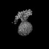 17356MC M: map data used to model this data C: citing same article ( |
|---|---|
| Similar structure data |
-Links
 PDBj
PDBj
- Assembly
| Deposited unit | 
|
|---|---|
| 1 |
|
-Components
| #1: Protein | Mass: 11150.034 Da / Num. of mol.: 1 Source method: isolated from a genetically manipulated source Source: (gene. exp.) Pseudomonas aeruginosa (bacteria) / Gene: ftsL, PA4419 / Production host: |
|---|---|
| #2: Protein | Mass: 43793.629 Da / Num. of mol.: 1 Source method: isolated from a genetically manipulated source Source: (gene. exp.) Pseudomonas aeruginosa (bacteria) / Gene: ftsW, PA4413 / Production host: References: UniProt: Q9HW00, peptidoglycan glycosyltransferase |
| #3: Protein | Mass: 62933.082 Da / Num. of mol.: 1 Source method: isolated from a genetically manipulated source Source: (gene. exp.) Pseudomonas aeruginosa (bacteria) / Gene: ftsI, pbpB, PA4418 / Production host: References: UniProt: G3XD46, serine-type D-Ala-D-Ala carboxypeptidase |
| #4: Protein | Mass: 10890.521 Da / Num. of mol.: 1 Source method: isolated from a genetically manipulated source Source: (gene. exp.) Pseudomonas aeruginosa (bacteria) / Gene: ftsB, PA3634 / Production host: |
| #5: Protein | Mass: 32290.223 Da / Num. of mol.: 1 Source method: isolated from a genetically manipulated source Source: (gene. exp.) Pseudomonas aeruginosa (bacteria) / Gene: ftsQ, PA4409 / Production host: |
| Has protein modification | N |
-Experimental details
-Experiment
| Experiment | Method: ELECTRON MICROSCOPY |
|---|---|
| EM experiment | Aggregation state: PARTICLE / 3D reconstruction method: single particle reconstruction |
- Sample preparation
| Component | Name: divisome complex FtsWIQLB / Type: COMPLEX / Entity ID: all / Source: RECOMBINANT |
|---|---|
| Source (natural) | Organism: |
| Source (recombinant) | Organism: |
| Buffer solution | pH: 7.8 |
| Specimen | Embedding applied: NO / Shadowing applied: NO / Staining applied: NO / Vitrification applied: YES |
| Vitrification | Cryogen name: ETHANE |
- Electron microscopy imaging
| Experimental equipment |  Model: Titan Krios / Image courtesy: FEI Company |
|---|---|
| Microscopy | Model: FEI TITAN KRIOS |
| Electron gun | Electron source:  FIELD EMISSION GUN / Accelerating voltage: 300 kV / Illumination mode: FLOOD BEAM FIELD EMISSION GUN / Accelerating voltage: 300 kV / Illumination mode: FLOOD BEAM |
| Electron lens | Mode: BRIGHT FIELD / Nominal defocus max: 2000 nm / Nominal defocus min: 1000 nm / Calibrated defocus min: 1000 nm / Calibrated defocus max: 2000 nm |
| Image recording | Electron dose: 50 e/Å2 / Film or detector model: FEI FALCON IV (4k x 4k) |
- Processing
| Software | Name: PHENIX / Version: 1.20.1_4487: / Classification: refinement | ||||||||||||||||||||||||
|---|---|---|---|---|---|---|---|---|---|---|---|---|---|---|---|---|---|---|---|---|---|---|---|---|---|
| EM software | Name: PHENIX / Category: model refinement | ||||||||||||||||||||||||
| CTF correction | Type: PHASE FLIPPING AND AMPLITUDE CORRECTION | ||||||||||||||||||||||||
| 3D reconstruction | Resolution: 3.3 Å / Resolution method: FSC 0.143 CUT-OFF / Num. of particles: 340758 / Symmetry type: POINT | ||||||||||||||||||||||||
| Refine LS restraints |
|
