[English] 日本語
Yorodumi
- PDB-8p05: Crystal structure of human Casein Kinase II subunit alpha (CK2a1)... -

+
Open data


ID or keywords:

Loading...

-
Basic information

Entry
Database: PDB / ID: 8p05
TitleCrystal structure of human Casein Kinase II subunit alpha (CK2a1) in complex with Leucettinib-92
ComponentsCasein kinase II subunit alpha
KeywordsTRANSFERASE / Kinase / CK2a / CSNK2A1 / Inhibitor / Leucettinib
Function / homology
Function and homology information


regulation of chromosome separation / positive regulation of aggrephagy / WNT mediated activation of DVL / Condensation of Prometaphase Chromosomes / protein kinase CK2 complex / symbiont-mediated disruption of host cell PML body / Receptor Mediated Mitophagy / Sin3-type complex / Synthesis of PC / RUNX1 interacts with co-factors whose precise effect on RUNX1 targets is not known ...regulation of chromosome separation / positive regulation of aggrephagy / WNT mediated activation of DVL / Condensation of Prometaphase Chromosomes / protein kinase CK2 complex / symbiont-mediated disruption of host cell PML body / Receptor Mediated Mitophagy / Sin3-type complex / Synthesis of PC / RUNX1 interacts with co-factors whose precise effect on RUNX1 targets is not known / Maturation of hRSV A proteins / negative regulation of apoptotic signaling pathway / positive regulation of Wnt signaling pathway / negative regulation of double-strand break repair via homologous recombination / chaperone-mediated protein folding / negative regulation of ubiquitin-dependent protein catabolic process / Signal transduction by L1 / peptidyl-threonine phosphorylation / Hsp90 protein binding / negative regulation of cysteine-type endopeptidase activity involved in apoptotic process / PML body / Wnt signaling pathway / Regulation of PTEN stability and activity / positive regulation of protein catabolic process / Cooperation of PDCL (PhLP1) and TRiC/CCT in G-protein beta folding / KEAP1-NFE2L2 pathway / double-strand break repair / rhythmic process / kinase activity / positive regulation of cell growth / peptidyl-serine phosphorylation / Regulation of TP53 Activity through Phosphorylation / protein stabilization / negative regulation of translation / non-specific serine/threonine protein kinase / regulation of cell cycle / cell cycle / protein phosphorylation / protein serine kinase activity / protein serine/threonine kinase activity / apoptotic process / DNA damage response / positive regulation of cell population proliferation / signal transduction / nucleoplasm / ATP binding / identical protein binding / nucleus / plasma membrane / cytosol
Similarity search - Function
Casein Kinase 2, subunit alpha / Serine/threonine-protein kinase, active site / Serine/Threonine protein kinases active-site signature. / Protein kinase domain / Serine/Threonine protein kinases, catalytic domain / Protein kinase, ATP binding site / Protein kinases ATP-binding region signature. / Protein kinase domain profile. / Protein kinase domain / Protein kinase-like domain superfamily
Similarity search - Domain/homology
Chem-WAK / Casein kinase II subunit alpha
Similarity search - Component
Biological speciesHomo sapiens (human)
MethodX-RAY DIFFRACTION / SYNCHROTRON / MOLECULAR REPLACEMENT / Resolution: 2.45 Å
AuthorsKraemer, A. / Meijer, L. / Knapp, S. / Structural Genomics Consortium (SGC)
Funding support1items
OrganizationGrant numberCountry
Not funded
CitationJournal: J.Med.Chem. / Year: 2023
Title: Leucettinibs, a Class of DYRK/CLK Kinase Inhibitors Inspired by the Marine Sponge Natural Product Leucettamine B.
Authors: Deau, E. / Lindberg, M.F. / Miege, F. / Roche, D. / George, N. / George, P. / Kramer, A. / Knapp, S. / Meijer, L.
History
DepositionMay 9, 2023Deposition site: PDBE / Processing site: PDBE
Revision 1.0May 17, 2023Provider: repository / Type: Initial release
Revision 1.1Nov 22, 2023Group: Data collection / Database references
Category: chem_comp_atom / chem_comp_bond ...chem_comp_atom / chem_comp_bond / citation / citation_author
Item: _citation.country / _citation.journal_abbrev ..._citation.country / _citation.journal_abbrev / _citation.journal_id_ASTM / _citation.journal_id_CSD / _citation.journal_id_ISSN / _citation.journal_volume / _citation.page_first / _citation.page_last / _citation.pdbx_database_id_DOI / _citation.pdbx_database_id_PubMed / _citation.title / _citation.year

-
Structure visualization

Structure viewerMolecule:
MolmilJmol/JSmol

Downloads & links

-
Assembly

Deposited unit
A: Casein kinase II subunit alpha
B: Casein kinase II subunit alpha
hetero molecules


Theoretical massNumber of molelcules
Total (without water)82,56616
Polymers80,6562
Non-polymers1,91014
Water63135
1
A: Casein kinase II subunit alpha
hetero molecules


Theoretical massNumber of molelcules
Total (without water)41,3799
Polymers40,3281
Non-polymers1,0518
Water181
TypeNameSymmetry operationNumber
identity operation1_555x,y,z1
Buried area1410 Å2
ΔGint-101 kcal/mol
Surface area15160 Å2
2
B: Casein kinase II subunit alpha
hetero molecules


Theoretical massNumber of molelcules
Total (without water)41,1877
Polymers40,3281
Non-polymers8596
Water181
TypeNameSymmetry operationNumber
identity operation1_555x,y,z1
Buried area940 Å2
ΔGint-66 kcal/mol
Surface area14900 Å2
Unit cell
Length a, b, c (Å)127.300, 127.300, 124.471
Angle α, β, γ (deg.)90.00, 90.00, 90.00
Int Tables number96
Space group name H-MP43212

-
Components

#1: Protein Casein kinase II subunit alpha / CK II alpha


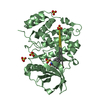
Mass: 40327.977 Da / Num. of mol.: 2
Source method: isolated from a genetically manipulated source
Source: (gene. exp.) Homo sapiens (human) / Gene: CSNK2A1, CK2A1 / Production host: Escherichia coli (E. coli)
References: UniProt: P68400, non-specific serine/threonine protein kinase
#2: Chemical
ChemComp-SO4 / SULFATE ION


Mass: 96.063 Da / Num. of mol.: 12 / Source method: obtained synthetically / Formula: SO4
#3: Chemical ChemComp-WAK / (4~{Z})-2-(1-adamantylamino)-4-(1,3-benzothiazol-6-ylmethylidene)-1~{H}-imidazol-5-one


Mass: 378.491 Da / Num. of mol.: 2 / Source method: obtained synthetically / Formula: C21H22N4OS / Feature type: SUBJECT OF INVESTIGATION
#4: Water ChemComp-HOH / water


Mass: 18.015 Da / Num. of mol.: 35 / Source method: isolated from a natural source / Formula: H2O
Has ligand of interestY

-
Experimental details

-
Experiment

ExperimentMethod: X-RAY DIFFRACTION / Number of used crystals: 1

-
Sample preparation

CrystalDensity Matthews: 3.13 Å3/Da / Density % sol: 60.65 %
Crystal growTemperature: 293 K / Method: vapor diffusion / pH: 5.5
Details: 0.2 M ammonia sulphate 0.1 M bis-tris pH 5.5 23-26% PEG 3350

-
Data collection

DiffractionMean temperature: 100 K / Serial crystal experiment: N
Diffraction sourceSource: SYNCHROTRON / Site: SLS / Beamline: X06SA / Wavelength: 0.999999 Å
DetectorType: DECTRIS EIGER X 16M / Detector: PIXEL / Date: May 6, 2023
RadiationProtocol: SINGLE WAVELENGTH / Monochromatic (M) / Laue (L): M / Scattering type: x-ray
Radiation wavelengthWavelength: 0.999999 Å / Relative weight: 1
ReflectionResolution: 2.45→45.05 Å / Num. obs: 38222 / % possible obs: 100 % / Redundancy: 13.4 % / CC1/2: 0.999 / Net I/σ(I): 15.1
Reflection shellResolution: 2.45→2.55 Å / Num. unique obs: 4227 / CC1/2: 0.858

-
Processing

Software
NameVersionClassification
REFMAC5.8.0352refinement
Aimlessdata scaling
XDSdata reduction
MOLREPphasing
RefinementMethod to determine structure: MOLECULAR REPLACEMENT / Resolution: 2.45→45.05 Å / Cor.coef. Fo:Fc: 0.954 / Cor.coef. Fo:Fc free: 0.923 / SU B: 11.019 / SU ML: 0.231 / Cross valid method: THROUGHOUT / ESU R: 0.328 / ESU R Free: 0.249 / Stereochemistry target values: MAXIMUM LIKELIHOOD / Details: HYDROGENS HAVE BEEN ADDED IN THE RIDING POSITIONS
RfactorNum. reflection% reflectionSelection details
Rfree0.25499 1922 5 %RANDOM
Rwork0.20883 ---
obs0.21122 36238 99.95 %-
Solvent computationIon probe radii: 0.8 Å / Shrinkage radii: 0.8 Å / VDW probe radii: 1.2 Å / Solvent model: MASK
Displacement parametersBiso mean: 60.478 Å2
Baniso -1Baniso -2Baniso -3
1-3 Å2-0 Å20 Å2
2--3 Å2-0 Å2
3----5.99 Å2
Refinement stepCycle: 1 / Resolution: 2.45→45.05 Å
ProteinNucleic acidLigandSolventTotal
Num. atoms5415 0 114 35 5564
Refine LS restraints
Refine-IDTypeDev idealDev ideal targetNumber
X-RAY DIFFRACTIONr_bond_refined_d0.0080.0125676
X-RAY DIFFRACTIONr_bond_other_d0.0010.0164971
X-RAY DIFFRACTIONr_angle_refined_deg1.5671.6557720
X-RAY DIFFRACTIONr_angle_other_deg0.561.58811545
X-RAY DIFFRACTIONr_dihedral_angle_1_deg6.845655
X-RAY DIFFRACTIONr_dihedral_angle_2_deg14.183539
X-RAY DIFFRACTIONr_dihedral_angle_3_deg17.40810918
X-RAY DIFFRACTIONr_dihedral_angle_4_deg
X-RAY DIFFRACTIONr_chiral_restr0.0780.2821
X-RAY DIFFRACTIONr_gen_planes_refined0.0080.026568
X-RAY DIFFRACTIONr_gen_planes_other0.0010.021192
X-RAY DIFFRACTIONr_nbd_refined
X-RAY DIFFRACTIONr_nbd_other
X-RAY DIFFRACTIONr_nbtor_refined
X-RAY DIFFRACTIONr_nbtor_other
X-RAY DIFFRACTIONr_xyhbond_nbd_refined
X-RAY DIFFRACTIONr_xyhbond_nbd_other
X-RAY DIFFRACTIONr_metal_ion_refined
X-RAY DIFFRACTIONr_metal_ion_other
X-RAY DIFFRACTIONr_symmetry_vdw_refined
X-RAY DIFFRACTIONr_symmetry_vdw_other
X-RAY DIFFRACTIONr_symmetry_hbond_refined
X-RAY DIFFRACTIONr_symmetry_hbond_other
X-RAY DIFFRACTIONr_symmetry_metal_ion_refined
X-RAY DIFFRACTIONr_symmetry_metal_ion_other
X-RAY DIFFRACTIONr_mcbond_it5.4576.262630
X-RAY DIFFRACTIONr_mcbond_other5.4566.262631
X-RAY DIFFRACTIONr_mcangle_it7.3889.3913281
X-RAY DIFFRACTIONr_mcangle_other7.3889.3913282
X-RAY DIFFRACTIONr_scbond_it6.7266.6823046
X-RAY DIFFRACTIONr_scbond_other6.4846.6543019
X-RAY DIFFRACTIONr_scangle_it
X-RAY DIFFRACTIONr_scangle_other8.7329.8234398
X-RAY DIFFRACTIONr_long_range_B_refined10.33279.8066211
X-RAY DIFFRACTIONr_long_range_B_other10.3479.7296199
X-RAY DIFFRACTIONr_rigid_bond_restr
X-RAY DIFFRACTIONr_sphericity_free
X-RAY DIFFRACTIONr_sphericity_bonded
LS refinement shellResolution: 2.45→2.514 Å / Total num. of bins used: 20
RfactorNum. reflection% reflection
Rfree0.416 122 -
Rwork0.367 2625 -
obs--99.85 %

+
About Yorodumi

-
News

-
Feb 9, 2022. New format data for meta-information of EMDB entries

New format data for meta-information of EMDB entries

  • Version 3 of the EMDB header file is now the official format.
  • The previous official version 1.9 will be removed from the archive.

Related info.:EMDB header

External links:wwPDB to switch to version 3 of the EMDB data model

-
Aug 12, 2020. Covid-19 info

Covid-19 info

URL: https://pdbjlvh1.pdbj.org/emnavi/covid19.php

New page: Covid-19 featured information page in EM Navigator.

Related info.:Covid-19 info / Mar 5, 2020. Novel coronavirus structure data

+
Mar 5, 2020. Novel coronavirus structure data

Novel coronavirus structure data

Related info.:Yorodumi Speices / Aug 12, 2020. Covid-19 info

External links:COVID-19 featured content - PDBj / Molecule of the Month (242):Coronavirus Proteases

+
Jan 31, 2019. EMDB accession codes are about to change! (news from PDBe EMDB page)

EMDB accession codes are about to change! (news from PDBe EMDB page)

  • The allocation of 4 digits for EMDB accession codes will soon come to an end. Whilst these codes will remain in use, new EMDB accession codes will include an additional digit and will expand incrementally as the available range of codes is exhausted. The current 4-digit format prefixed with “EMD-” (i.e. EMD-XXXX) will advance to a 5-digit format (i.e. EMD-XXXXX), and so on. It is currently estimated that the 4-digit codes will be depleted around Spring 2019, at which point the 5-digit format will come into force.
  • The EM Navigator/Yorodumi systems omit the EMD- prefix.

Related info.:Q: What is EMD? / ID/Accession-code notation in Yorodumi/EM Navigator

External links:EMDB Accession Codes are Changing Soon! / Contact to PDBj

+
Jul 12, 2017. Major update of PDB

Major update of PDB

  • wwPDB released updated PDB data conforming to the new PDBx/mmCIF dictionary.
  • This is a major update changing the version number from 4 to 5, and with Remediation, in which all the entries are updated.
  • In this update, many items about electron microscopy experimental information are reorganized (e.g. em_software).
  • Now, EM Navigator and Yorodumi are based on the updated data.

External links:wwPDB Remediation / Enriched Model Files Conforming to OneDep Data Standards Now Available in the PDB FTP Archive

-
Yorodumi

Thousand views of thousand structures

  • Yorodumi is a browser for structure data from EMDB, PDB, SASBDB, etc.
  • This page is also the successor to EM Navigator detail page, and also detail information page/front-end page for Omokage search.
  • The word "yorodu" (or yorozu) is an old Japanese word meaning "ten thousand". "mi" (miru) is to see.

Related info.:EMDB / PDB / SASBDB / Comparison of 3 databanks / Yorodumi Search / Aug 31, 2016. New EM Navigator & Yorodumi / Yorodumi Papers / Jmol/JSmol / Function and homology information / Changes in new EM Navigator and Yorodumi

Read more